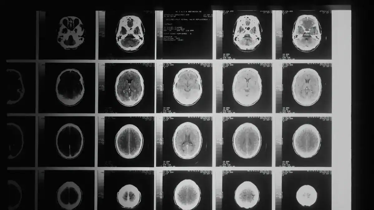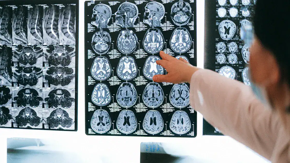
A brain bleed, or intracranial hemorrhage, occurs when blood escapes from a vessel inside the skull. This brain hemorrhage is severe, rapidly depriving brain cells of oxygen.
Cerebral bleeding affects approximately 2.5 out of every 10,000 individuals annually, with the incidence of intracerebral hemorrhage being 24.6 cases per 100,000 person-years. Brain bleeds, including intracerebral hemorrhage, are a type of stroke. Prompt medical treatment is critical for recovery. The journey to brain bleed recovery after a brain hemorrhage is often long and challenging. This article outlines a healing timeline, detailing what patients and families can expect regarding brain recovery.
Key Takeaways
Brain bleed recovery happens in stages. These stages are acute, subacute, and chronic. Each stage has different healing goals.
Many things affect recovery. These include the bleed’s size and location. Quick medical help and the patient’s age also matter.
Rehabilitation is very important. Physical, occupational, and speech therapies help patients get back lost skills. Cognitive therapy also helps the brain.
Daily habits support brain healing. Get enough rest and sleep. Eat healthy foods and drink water. Take medicines as told. Manage stress well.
Understanding Critical Recovery Stages

Recovery from a brain hemorrhage unfolds in distinct stages. Each stage presents unique challenges and opportunities for healing. Understanding these brain bleed recovery stages helps patients and their families prepare for the journey ahead. The overall recovery duration varies greatly among individuals.
Acute Phase: Immediate Intervention
The acute stage of brain bleed recovery typically lasts for the first few days after the event. During this period, medical professionals focus on stabilizing the patient’s condition. They address immediate health threats, control bleeding, and alleviate pressure in the brain. This stage is crucial for initiating the healing process. Doctors implement several critical interventions.
They administer anticonvulsants to prevent seizure recurrence. Antihypertensive agents reduce blood pressure. Osmotic diuretics decrease intracranial pressure. Medical teams also stabilize vital signs. They may perform endotracheal intubation for decreased consciousness and poor airway protection. Intubation and hyperventilation help if intracranial pressure is elevated.
Administration of mannitol controls intracranial pressure. Monitoring glucose levels ensures normoglycemia. Antacids prevent gastric ulcers. In some cases, evacuation of the hematoma occurs via open craniotomy or endoscopy. This offers a promising ultra-early-stage treatment. Other interventions include reversal of anticoagulation, modest blood pressure reduction, and monitoring of temperature. Intracranial pressure monitoring and management, hematoma evacuation, and external ventricular drainage are also performed in selected patients. Airway protection remains a top priority. This initial period of critical recovery sets the foundation for future healing of the brain.
Subacute Phase: Early Rehabilitation
Following the acute phase, patients enter the subacute phase. This stage typically spans the first few weeks to six months after the brain hemorrhage. During this time, the majority of significant recovery often happens. The brain begins to repair itself, and patients start early rehabilitation efforts.
For mild cases of a bleed, individuals might see substantial improvement within approximately three weeks. Moderate cases often show significant progress in about six weeks.
Rehabilitation focuses on regaining lost functions. This includes physical, occupational, and speech therapies. The brain’s plasticity allows for remarkable adaptation during this period. Patients work to relearn daily activities and improve their motor skills. The recovery time in this phase is vital for maximizing functional independence.
Chronic Phase: Long-Term Adaptation
The chronic phase of brain bleed recovery extends beyond the first six months. It can continue for several years. During this stage, patients continue to improve and adapt to any remaining disabilities.
Rehabilitation efforts continue, focusing on improving quality of life and maximizing independence. This may involve advanced therapies such as constraint-induced movement therapy, mirror therapy, or repetitive task training. The goal is to help the patient adapt to life after stroke, manage any long-term disabilities, and prevent recurrent strokes. This stage underscores the importance of ongoing support and resources for stroke survivors.
Gradual improvements may continue for up to two years, especially if the patient continues with physical and occupational therapies.
Improvements slow down substantially after two years but may still occur many years after injury. This long-term recovery duration highlights the brain’s incredible capacity for healing. Studies show that 50-64% of survivors achieve independence in daily activities between 6 to 32 months post-intracerebral hemorrhage. Achieving full recovery from a brain bleed is a long process. It requires consistent effort and support. This cerebral hemorrhage recovery journey emphasizes the resilience of the human brain.
Factors Influencing Brain Bleed Recovery
Many factors affect a person’s recovery from a brain hemorrhage. These elements can significantly impact the healing timeline and the extent of functional improvement. Understanding these factors helps patients and their families set realistic expectations for brain bleed recovery.
Hemorrhage Type, Size, Location
The specific characteristics of the brain hemorrhage play a crucial role in recovery. Different types of bleeds affect the brain in distinct ways. For example, an intracerebral hemorrhage (ICH) occurs within the brain tissue itself. Aneurysmal subarachnoid hemorrhage (SAH) involves bleeding into the space surrounding the brain.
A concomitant intracerebral hemorrhage (ICH) is a poor prognostic factor for an unfavorable outcome following aneurysmal subarachnoid hemorrhage (SAH). This means a person with both types of bleeds faces a tougher recovery.
The size of the bleed also matters greatly. Larger bleeds often cause more extensive brain damage after a brain bleed. This leads to more severe neurological deficits. The location of the hemorrhage within the brain is equally important.
A bleed in a critical area, such as the brainstem, can have more devastating effects than one in a less vital region. While intraparenchymal hematoma was frequently observed in one study, it did not significantly affect mortality or unfavorable outcomes. This lack of impact was potentially due to a high rate of hematoma evacuation surgery. Doctors performed this surgery in 82% of affected patients. This may have reduced the risk of secondary brain injury.
Speed of Medical Treatment
Prompt medical treatment is paramount for improving outcomes after a brain hemorrhage. The faster doctors intervene, the better the chances for a positive recovery. Early and aggressive treatment plans are crucial.
They focus on neurological monitoring, managing elevated intracranial pressure or cerebral ischemia, and preventing secondary medical complications. Key interventions include identifying the underlying cause of hemorrhage, correcting elevated blood pressure, reversing coagulopathy, and providing supportive care in a neurological ICU.
Microsurgical intervention for hemorrhagic brainstem cavernous malformations (BSCMs) that exceeds 42 days after a prior hemorrhage is associated with an increased risk of unfavorable neurological outcomes.
The ideal window for microsurgical treatment of BSCM is approximately within 42 days following the hemorrhage onset. Treatment within 21 days after hemorrhage onset showed a progressive increase in the odds of unfavorable outcomes. However, it remained beneficial for favorable outcomes until 42 days. Beyond 42 days, the odds of unfavorable outcomes increased. Subacute intervention (3-8 weeks) might offer protective effects against re-hemorrhage and lower cranial nerve deficits compared to acute intervention (within 3 weeks).
Prompt surgical removal of a brain hemorrhage also significantly improves patient outcomes. This involves clearing 70% or more of the clot to leave no more than 15 milliliters. This approach uses a minimally invasive procedure with a cannula to aspirate clotted blood. Doctors then administer alteplase (Activase®) through a catheter to dissolve remaining clots. The MISTIE III trial showed that achieving these volume-reduction goals is critical for the surgery’s effectiveness. Each additional milliliter of clot removed improved the odds of a good outcome by 10%.
Specific medical interventions are most effective when administered promptly:
Airway, Breathing, and Blood Pressure Management: Doctors first assess and manage the patient’s airway, breathing, blood pressure, and signs of increased intracranial pressure (ICP). Intubation may be necessary for aspiration risk, ventilatory failure, or elevated ICP.
Intracranial Pressure (ICP) Control: For stuporous or comatose patients, or those with signs of brainstem herniation, emergency measures include elevating the head to 30 degrees. Doctors also rapidly infuse 1.0–1.5 g/kg of 20% mannitol. Hyperventilation to a pCO2 of 30–35 mmHg helps. These actions aim to quickly lower ICP before definitive neurosurgical procedures.
Blood Pressure Regulation: Elevated blood pressure is common in ICH patients. Guidelines recommend maintaining a mean arterial pressure below 130 mmHg for patients with a history of hypertension. For those with elevated ICP and an ICP monitor, cerebral perfusion pressure (MAP–ICP) should be kept above 70 mmHg.
Early Hemostatic Therapies: Prompt identification and reversal of effects from thrombolytic, antiplatelet, or anticoagulant use are crucial. This involves blood tests and making blood products available for urgent administration. Specific reversal agents like protamine sulfate for heparin and vitamin K with FFP/clotting factor concentrates for warfarin are recommended.
Patient Age and Health
A patient’s age significantly influences the brain’s capacity for recovery. Younger individuals generally possess more robust plasticity mechanisms. These include synaptogenesis, dendritic remodeling, and neurogenesis. This leads to faster and more complete functional recovery.
Older individuals exhibit reduced plastic potential. Factors like cellular senescence, diminished trophic support, chronic inflammation, and impaired neural stem cell function contribute to this. Their brains show a prolonged and heightened inflammatory gene expression profile. They also have reduced expression of genes critical for repair and plasticity. This leads to prolonged recovery and cognitive deterioration.
A rat model of focal ischemia showed that younger rats achieved faster and more complete functional recovery compared to older groups. This indicates more pronounced neuroplasticity in young brains.
Aging also brings other challenges:
Gene Expression: Ischemic stroke triggers age-modulated gene expression changes. Young brains show an acute upregulation of stress and inflammatory genes followed by reparative genes. Aged brains exhibit prolonged inflammation and diminished induction of repair/plasticity genes.
Cellular and Molecular Changes: Aging leads to a decline in mitochondrial efficiency, increased reactive oxygen species (ROS), reduced ATP, and heightened metabolic stress. These impair neuronal recovery.
Vascular Changes: The aging brain experiences significant vascular changes. These include blood-brain barrier (BBB) breakdown. This increases permeability to neurotoxic substances and inflammatory mediators, exacerbating damage.
Complications and Co-morbidities
Pre-existing health conditions, or co-morbidities, can complicate brain bleed recovery. These conditions often lead to poorer outcomes. A study found that current smoking was the most common risk factor (29%). Hypertension (17%), diabetes (8%), and hyperlipidemia (6%) followed. Smoking was consistently associated with worse scores across all four outcome indices. Hypertension and diabetes were linked to worse scores on the Rivermead Post-Concussion Symptoms Questionnaire (RPQ). Hypertension also showed an association with worse scores on the 18-Item Brief Symptom Inventory Global Severity Index (BSI-18-GSI). Individuals with one or more vascular risk factors generally experienced worse outcomes compared to those with none.
Pre-injury vascular risk factors, especially smoking, are associated with poorer outcomes following Traumatic Brain Injury (TBI). Aggressive post-injury management of these vascular risk factors could improve TBI recovery.
Medical comorbidities and complications after a brain bleed also influence the Functional Independence Measure (FIM) change and the length of stay during inpatient rehabilitation for traumatic brain injury (TBI) patients.
In a study evaluating early rehabilitation for patients with moderate and severe traumatic brain injury, comorbidities such as hypertension, diabetes mellitus, and asthma were registered as part of the patient evaluation. These conditions can increase the chances of a second brain hemorrhage or lead to permanent neurological damage. They also require more intensive and longer rehabilitation periods. Recognizing signs of complications early helps manage these challenges effectively.
Rehabilitation for Brain Hemorrhage Healing

Rehabilitation plays a vital role in recovery after a brain hemorrhage. It helps patients regain lost abilities and improve their quality of life. This process involves various specialized therapies. These therapies work to retrain the brain and body.
Physical Therapy
Physical therapy helps patients improve motor function and reduce disability after a brain hemorrhage. Therapists aim to increase physical activity and fitness levels. They also work to improve overall quality of life. This therapy facilitates structural brain remodeling. Active rehabilitation protocols, including gradual exercise, promote neuroplasticity. This helps the brain adapt to new demands. Physical therapy often involves three stages. First, therapists prepare the brain with cardio and breathing exercises. This boosts blood flow and releases neurochemicals. Second, they activate the brain with neuromuscular and sensorimotor therapies. Third, patients recover and rest with massage and mindfulness techniques. Studies show physical activities like treadmill running improve motor function after intracerebral hemorrhage. Early constraint therapy also helps recovery in models of subcortical hemorrhage. These interventions harness the brain’s ability to rewire itself.
Occupational Therapy
Occupational therapists (OTs) help patients regain independence in daily living activities. This includes tasks like dressing, cooking, and personal hygiene. OTs use tailored strategies and adaptive techniques. They help patients overcome physical and cognitive limits.
Therapy sessions involve practicing daily tasks and using special equipment. Occupational therapy significantly helps patients achieve maximum function. Patients become more independent in personal activities like feeding and bathing. OTs teach specific techniques, such as dressing the affected side first for individuals with hemiplegia. They recommend adaptive equipment like sock aides or raised toilet seats. OTs also provide cognitive adaptations, such as using checklists or minimizing distractions. This consistent practice stimulates neuroplasticity, helping the brain relearn skills.
Speech and Language Therapy
A brain hemorrhage can cause communication problems. Aphasia is common, affecting speech and language. Types include expressive aphasia, where people struggle to speak, and receptive aphasia, where they have trouble understanding. Global aphasia is severe, affecting all language skills. Speech and language therapy aims to restore these skills. Therapists use language exercises to improve comprehension and vocabulary. They also use cognitive therapy to boost memory and attention. Speech production strategies strengthen speech muscles. Swallowing therapy helps with eating and drinking safely. Augmentative and alternative communication (AAC) systems offer tools like communication boards. This comprehensive treatment helps patients regain functional communication.
Cognitive Rehabilitation
A brain bleed often affects cognitive functions. These include non-verbal IQ, processing speed, and executive function. Language and memory can also suffer. Cognitive rehabilitation helps patients improve these areas. Therapists teach compensatory strategies, like using a memory notebook or daily planner. They also use task analysis, breaking down tasks into smaller steps. Attention-enhancing exercises improve information retention.
Exercises increase capacity for attention and working memory. Cognitive rehabilitation uses both restorative and compensatory approaches. Restorative methods involve repeated exercises to rebuild skills. Compensatory methods teach ways to bypass impaired functions, such as using alarms. Problem-solving exercises like Sudoku and chess also stimulate the brain.
Practical Strategies for Brain Recovery
Implementing practical strategies significantly supports the brain’s healing process. These daily practices help manage symptoms and promote long-term well-being after a brain hemorrhage.
Rest and Sleep
Adequate rest and sleep are crucial for brain healing. The brain needs proper rest after a brain hemorrhage. Establish a consistent sleep schedule. Go to bed and wake up at the same time daily, even on weekends. Keep daytime naps under 30 minutes. Many brain injury survivors experience insomnia. This highlights good sleep hygiene’s importance.
Avoid electronics and bright lights before bedtime. Consider cognitive-behavioral techniques for persistent sleep difficulties. Choose medications with fewer cognitive side effects, like trazadone, if needed. Make your bedtime routine calming. Engage in activities like stretching or listening to music. Ensure your diet supports neurotransmitter production with magnesium and good fatty acids. Avoid high sugar or trans fat diets. These diets link to poor sleep quality.
Nutrition and Hydration
Proper nutrition and hydration significantly help brain bleed recovery. Certain foods support brain health during recovery from a brain hemorrhage. Berries, especially blueberries, improve memory and cognitive functions. Eggs and avocados offer cognitive boosts.
Meat provides protein and zinc. Zinc is crucial for the immune system and memory. Walnuts are rich in omega-3s. Turmeric promotes neuronal repair. Avoid foods high in saturated fat and processed sugar. These reduce neuroplasticity. The Mediterranean diet, rich in fish, fruits, and vegetables, benefits brain injury recovery. Always consult a physician or dietitian before starting new diets. Incorporate fermented foods like sauerkraut and yogurt. Maintain a consistent eating schedule. Eat every four to five hours.
Medication Adherence
Adhering to prescribed medications is vital for healing after a brain hemorrhage. Doctors prescribe these to manage symptoms like pain or seizures. Following the schedule helps prevent complications and supports the brain’s healing. Never adjust dosages or stop medications without consulting your doctor. This consistent intake is a key part of the overall treatment plan for a brain bleed. This treatment helps manage the effects of the hemorrhage.
Stress Management
Managing stress is another key strategy for brain recovery after a hemorrhage. Survivors can reduce sensory overload by eating simple foods. They should monitor pain, stress, and fatigue levels. Taking regular brain-recharging breaks helps. Go to a quiet place, close eyes, and breathe deeply. Caregivers can support survivors by asking about sensory overload triggers.
They should create non-overwhelming social environments. Counseling and therapy, especially CBT, help manage emotional responses. Relaxation techniques like mindfulness reduce stress. Establish structured daily activities for stability. Peer support groups offer belonging. Caregivers should establish structured routines and recognize triggers for frustration. Seek professional guidance from neuropsychologists for tailored strategies after a hemorrhage. This helps with the long-term effects of the bleed.
Brain bleed recovery involves acute, subacute, and chronic stages, requiring a multidisciplinary approach for optimal healing. While recovery from a brain hemorrhage presents challenges, consistent rehabilitation and adherence to medical advice significantly improve outcomes.
Improvements can continue for up to two years, with some individuals showing continued functional recovery even after five years. The brain demonstrates remarkable resilience and adaptation, allowing for meaningful recovery and improved quality of life, even when facing long-term physical or cognitive challenges from a brain hemorrhage. Patients often report their functional status as “normal for my age,” despite physical limitations. However, brain bleeds can lead to persistent consequences, impacting employment and social activities.
FAQ
What is the typical recovery time for a brain bleed?
Recovery time varies greatly. Mild cases often show significant improvement within three weeks. Moderate cases may take about six weeks. Most major recovery happens within the first six months. Smaller improvements can continue for up to two years.
What are common long-term effects of a brain bleed?
Long-term effects can include physical weakness, speech difficulties, and cognitive changes. Patients may experience memory problems or trouble with executive functions. Rehabilitation helps manage these challenges. Many survivors achieve independence in daily activities.
What role does diet play in brain bleed recovery?
A healthy diet supports brain healing. Foods rich in omega-3s, antioxidants, and essential nutrients are beneficial. Berries, walnuts, and lean proteins help. Avoiding processed foods and excessive sugar is also important. Proper nutrition aids overall brain health.
What is neuroplasticity and how does it help recovery?
Neuroplasticity is the brain’s ability to reorganize itself. It forms new neural connections throughout life. After a brain bleed, neuroplasticity allows the brain to adapt and relearn lost functions. Rehabilitation therapies actively stimulate this process, promoting healing.


