Arm anatomy is the study of the structural components that allow the arm to perform its many functions. These components include bones, muscles, and nerves. Understanding these interconnected systems is crucial for comprehending movement, strength, and sensation in the arm.
The coordinated effort of these parts enables the arm’s remarkable capabilities. This blog post provides a clear, systematic overview of these essential anatomical structures.
Key Takeaways
The arm has three main parts: the upper arm, forearm, and hand. Each part has special bones, muscles, and nerves that help it move.
Arm bones like the humerus, radius, and ulna give the arm its shape and allow movement. Joints like the shoulder and elbow help the arm bend and twist.
Muscles in the arm, such as the biceps and triceps, pull on bones to make the arm move. They help with lifting, throwing, and writing.
Nerves in the arm send messages between the brain and muscles. This helps you move your arm and feel things like touch and temperature.
Bones, muscles, and nerves work together perfectly. This teamwork allows the arm to do many different things, from strong lifts to small, careful movements.
Arm Anatomy Explained: An Overview
Image Source: unsplash
Defining the Arm (Upper Extremity)
The arm, also known as the upper extremity, forms a highly functional and adaptable part of the human body. It consists of three primary segments. These segments are the upper arm, the forearm, and the hand. The upper arm extends from the shoulder to the elbow. It houses a single, robust bone. The forearm then continues from the elbow to the wrist.
It contains two distinct bones that allow for rotation. Finally, the hand, a complex structure, includes the wrist, the palm, and the fingers. Each segment possesses unique anatomical features. These features contribute to the arm’s overall versatility. The study of this intricate structure is known as anatomy. The joints connecting these segments, such as the shoulder, elbow, and wrist, provide crucial mobility. This integrated design allows for a wide spectrum of physical interactions.
Functional Importance of Arm Structure
The arm’s intricate structure underpins its profound functional importance. It enables a vast and diverse range of movements. Individuals rely on their arms for gross motor activities like lifting heavy objects or throwing a ball. They also use them for fine motor tasks, such as writing, buttoning a shirt, or playing a musical instrument.
The powerful muscles, anchored to the bones, generate the necessary strength and precision for these actions. Furthermore, a sophisticated network of nerves permeates the entire arm.
These nerves transmit vital signals. They control muscle contractions and convey sensory information back to the brain. This sensory input allows individuals to perceive touch, temperature, and pressure.
Arm Bones: The Skeletal Structure
The human arm contains a complex framework of bones. Each upper limb contains 30 bones. These bones provide support, allow movement, and protect vital structures. They form the foundation for the arm’s remarkable capabilities.
Upper Arm Bone: The Humerus
The humerus is the single bone of the upper arm. It extends from the shoulder to the elbow. This long bone connects to the shoulder blade at the top and the forearm bones at the bottom. The humerus plays a crucial role in arm movement and strength.
The humerus, as the single bone of the upper arm, can be vulnerable to injury. Falls often cause fractures, especially onto an outstretched arm. Accidents or injuries that apply direct force to the shoulder and upper arm, like car crashes, also lead to breaks. Repetitive stress from activities such as weightlifting can strain the bone. Underlying conditions, including metabolic bone disease or osteoporosis, weaken the bones. Bone tumors can also compromise the strength of this single bone of the upper arm. Common fracture sites include the proximal humerus, which is the top end of the bone.
Forearm Bones: Radius and Ulna
The forearm contains two distinct bones: the radius and the ulna. These are the paired bones of the forearm. The ulna sits medially, closer to the body’s midline. The radius is positioned laterally, on the thumb side. These two bones work together to allow complex forearm movements.
The ulna is generally longer than the radius.
Bone
Sex
Mean Length (mm)
SD Length (mm)
Mean Volume (cm³)
SD Volume (cm³)
Radius
Female
210.65
11.50
31.95
5.99
Radius
Male
228.59
11.19
45.99
6.83
Ulna
Female
227.80
11.73
37.30
6.49
Ulna
Male
245.23
10.92
53.23
6.89
The radius rotates around the ulnar head. This action achieves forearm rotation. This rotation happens along a longitudinal axis. This axis extends from the center of the radial head at the Proximal Radioulnar Joint (PRUJ) to the fovea of the ulnar head at the wrist. The PRUJ is a ball-and-socket joint. The radial head, with its convex articulating surface, fits into the radial notch of the ulna. The annular ligament encloses it.
At the Distal Radioulnar Joint (DRUJ), the ulnar head moves in a rolling and sliding motion. It moves within the sigmoid notch of the radius. Ligaments maintain the stability of these joints. These ligaments include the dorsal and palmar radioulnar ligaments and the interosseous membrane (IOM). The IOM links both bones. It prevents proximal migration of the radius. Its central band provides significant longitudinal stiffness.
Key Arm Joints
The arm features several key joints. These joints allow for a wide range of motion. The shoulder joint connects the humerus to the shoulder blade. The elbow joint connects the humerus to the radius and ulna. The wrist joint connects the forearm bones to the hand bones.
The elbow joint relies on strong ligaments for stability.
Annular Ligament (AL): This ligament surrounds the head of the radius. It helps maintain contact between the radius and humerus bones.
Medial Collateral Ligament (MCL): This ligament has anterior and posterior bands. It originates at the medial epicondyle and attaches to the ulna.
Lateral Collateral Ligament (LCL): This short band extends from the lateral epicondyle to the annular ligament.
Radial collateral ligament: It extends from the lateral epicondyle of the humerus. It merges with the annular ligament of the radius. This ligament encircles the radial head.
Ulnar collateral ligament: It originates from the medial epicondyle. It attaches to both the coronoid process and olecranon of the ulna.
The shoulder joint is highly mobile but also prone to various issues.
Articular pathologies: These include osteoarthritis of the glenohumeral joint and acromioclavicular joint.
Bone pathologies: Tumors, avascular necrosis, and fractures can affect the bones.
Soft tissue local pathologies: Rotator cuff tendinopathy, biceps tendinopathy, and shoulder instability are common.
Inflammatory rheumatic diseases: Polymyalgia rheumatica and rheumatoid arthritis can impact the joint.
Systemic diseases: Malignancy and infection also cause problems.
Strains and Sprains: These involve stretching or tearing of ligaments or muscles.
Shoulder Instability: This chronic condition leads to frequent dislocations.
Rotator Cuff Tear: This is an injury to the tendons supporting the shoulder joint.
Shoulder Bursitis: This involves inflammation of the bursa.
Shoulder Impingement: This occurs when tendons become inflamed due to repeated overhead movements.
Frozen Shoulder (Adhesive Capsulitis): This causes pain and stiffness.
Bicep Tendon Rupture: This is a tear of the biceps tendon.
These bones and joints form the essential skeletal structure of the arm. They provide the framework for all arm movements.
Arm Muscles: Movement & Strength
The arm’s ability to move and exert force comes from its many muscles. These muscles attach to the bones. They pull on the bones to create movement. The humerus, the upper arm bone, connects to 13 different muscles. These muscles work together. They allow a wide range of arm movements. Understanding these muscles helps explain the arm’s strength and flexibility.
Upper Arm Muscles and Functions
The upper arm contains two main groups of muscles: anterior and posterior. The anterior compartment muscles primarily bend the elbow. The posterior compartment muscles straighten the elbow.
The anterior compartment includes three key muscles:
Biceps Brachii: This prominent muscle has two heads. It flexes the elbow and supinates the forearm.
Part of Biceps Brachii
Origin
Insertion
Long Head
Supraglenoid tubercle of the scapula
Radial tuberosity of the radius
Short Head
Apex of the coracoid process of the scapula
Radial tuberosity of the radius
Common Insertion
Deep fascia of the forearm (via bicipital aponeurosis)
Brachialis: This muscle lies deep to the biceps. It is the primary flexor of the elbow.
Muscle Head
Origin
Insertion
Superficial Head
Anterior surface of the humerus, distal to the deltoid tuberosity.
Coronoid process and tuberosity of the ulna.
Deep Head
Anterior surface of the humerus, distal to the deltoid tuberosity.
Coronoid process and tuberosity of the ulna.
Coracobrachialis: This muscle helps flex and adduct the arm at the shoulder.
The posterior compartment contains one large muscle:
Triceps Brachii: This muscle has three heads. It extends the elbow, straightening the arm.
The deltoid muscle, though part of the shoulder, plays a crucial role in arm movement. It covers the shoulder joint.
Overall Function: It helps move shoulders and arms.
Anterior Section: This section lifts arms in front (shoulder flexion). It assists in crossing the arm over the chest (transverse flexion). It also helps lift the arm up and out to the side (shoulder abduction). It rotates the humerus.
Middle Section: This section lifts arms out to the side (shoulder abduction).
Posterior Section: This section helps extend the shoulder back behind the body. It can also help rotate the humerus.
Forearm Muscles and Compartments
The forearm also has anterior and posterior compartments. These compartments contain many muscles. They control wrist, hand, and finger movements.
The anterior compartment muscles primarily flex the wrist and fingers. They also pronate the forearm.
Level
Muscle
Superficial
Flexor carpi radialis
Superficial
Palmaris longus
Superficial
Flexor carpi ulnaris
Superficial
Pronator teres
Superficial (or Intermediate)
Flexor digitorum superficialis
Deep
Flexor digitorum profundus
Deep
Flexor pollicis longus
Deep
Pronator quadratus
The pronator teres muscle primarily performs pronation of the forearm. This action turns the palm backward from the anatomical position. It also assists in the flexion of the forearm.
The posterior compartment muscles primarily extend the wrist and fingers. They also supinate the forearm. These muscles are generally smaller than the anterior compartment muscles. They allow for fine motor control.
Muscle Actions and Attachments
Muscles create movement by contracting. They pull on the bones to which they attach. Each muscle has an origin and an insertion. The origin is the stable attachment point. The insertion is the movable attachment point. When a muscle contracts, the insertion moves towards the origin.
For example, the biceps brachii muscle originates on the scapula. It inserts on the radius. When it contracts, it pulls the radius towards the scapula. This action bends the elbow. The brachialis muscle also contributes significantly to this bending motion.
The coordinated action of these many muscles allows for the arm’s incredible range of motion. They enable everything from powerful lifting to delicate finger movements. Understanding these attachments and actions helps explain how the arm performs its diverse functions.
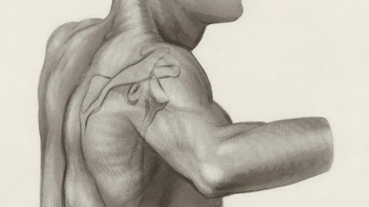
Arm Nerves: The Brachial Plexus & Beyond
Image Source: unsplash
The arm’s intricate movements and sensations rely on a complex network of nerves. These nerves transmit signals between the brain, spinal cord, and the arm’s muscles and skin. Understanding this neural pathway is crucial for comprehending how the arm functions.
The Brachial Plexus Origin and Network
The brachial plexus forms a critical network of nerves. It originates from the anterior primary rami of spinal nerves C5, C6, C7, C8, and T1. These roots are the anterior primary rami of the spinal nerves after they supply the neck muscles.
In some cases, prefixed or postfixed formations can involve C4 or T2, respectively. This network then branches out, innervating every structure of the arm. The brachial plexus ensures the arm receives both motor commands for movement and sensory information from its various parts.
The brachial plexus gives rise to five major terminal branches. These branches distribute throughout the arm, forearm, and hand.
Musculocutaneous nerve
Axillary nerve
Radial nerve
Median nerve
Ulnar nerve
These five main nerves control specific muscle groups and provide sensation to distinct skin areas.
Major Nerves of the Upper Arm
Several important nerves travel through the upper arm. Each nerve has specific motor and sensory roles.
The musculocutaneous nerve is one such important nerve. It primarily controls muscles in the anterior compartment of the upper arm. This nerve innervates the following muscles:
Biceps brachii
Brachialis
Coracobrachialis
These muscles help flex the elbow and supinate the forearm. The musculocutaneous nerve also provides sensory input to the lateral forearm skin.
The radial nerve is another significant nerve in the upper arm. It travels around the back of the humerus. This nerve primarily controls the triceps muscle, which extends the elbow. The radial nerve also provides sensory innervation to specific areas.
It supplies the lower outer aspect and posterior surface of the arm. Specifically, the lower lateral cutaneous nerve of the arm innervates the lateral aspect of the arm, inferior to the deltoid muscle’s insertion. The posterior cutaneous nerve of the arm provides sensation to the posterior surface of the arm. This sensory distribution allows individuals to feel touch, pain, and temperature in these regions.
The forearm also contains major nerves that extend into the hand. These nerves control fine motor skills and provide detailed sensory feedback.
The median nerve travels down the middle of the forearm. It provides motor supply to most of the flexor muscles in the forearm. Exceptions include the flexor carpi ulnaris and the ulnar head of the flexor digitorum profundus. The median nerve’s motor functions in the forearm include:
Bending and straightening the wrists, thumbs, and first three fingers.
Rotating the forearm and hand to turn the palm downward.
Its deep (volar interosseous) branch controls deeper muscles in the front part of the forearm. Its muscular branch controls movement in the forearm’s superficial muscles, close to the skin.
The ulnar nerve runs along the medial side of the forearm. It provides motor control to some forearm muscles and many small hand muscles. The ulnar nerve also supplies sensory input to specific hand areas. It provides sensory innervation to:
The fifth digit.
The medial half of the fourth digit.
The corresponding part of the palm.
This sensory input comes via its palmar cutaneous branch, dorsal cutaneous branch, and superficial branch. The palmar cutaneous branch innervates the medial half of the palm.
The dorsal cutaneous branch innervates the dorsal surface of the medial one and a half fingers and the associated dorsal hand area. The superficial branch innervates the palmar surface of the medial one and a half fingers. Overall, it supplies the medial one and a half fingers and the associated palm area. These nerves are essential for the arm’s complex functions.
Arm Anatomy: Interconnected Systems
Bone-Muscle-Nerve Coordination
The arm’s remarkable abilities stem from the precise coordination of its bones, muscles, and nerves. These systems do not work alone. They form a unified network.
Nerves send signals to muscle, telling it to contract. Muscles then pull on bones, creating movement. This process also involves constant feedback. Proprioceptors, special sensory receptors in muscles and joints, provide essential information.
Muscle spindles tell the brain about muscle stretch. Golgi tendon organs report on tendon force. Joint receptors become active when joint angles reach their limits. Cutaneous receptors detect skin stretch from joint movements. This integrated feedback helps the brain understand the arm’s position and movement.
Motor units also play a key role in this coordination. A motor unit includes a motor neuron and the muscle fibers it controls. For smooth arm movements, motor units fire asynchronously. This means they contract one after another, not all at once. This asynchronous firing ensures a smooth, controlled muscle contraction.
It prevents jerky movements. When a person lifts a light object, the body recruits small motor units first. For heavier objects, it recruits larger motor units. This distributed use of muscles allows for precise control over strength. It also helps prevent early fatigue. This complex interplay of bones, muscles, and nerves makes every arm action possible.
Functional Significance of Integration
The integration of bones, muscles, and nerves gives the arm its incredible functional range. This includes everything from powerful lifting to delicate fine motor skills.
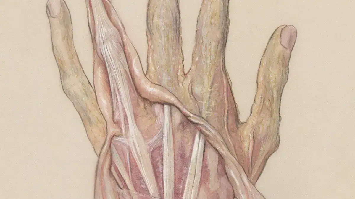
The hand, for example, shows this integration perfectly. Its skeletal structure, with many small bones, provides support. Soft tissues like ligaments and tendons connect these bones and muscles. Nerves in the hand provide sensation and control the muscles.
This collaboration enables precise movements. For instance, stabilizing the pinky and ring fingers allows for more accurate use of the pointer and middle fingers. Intrinsic hand muscles define arches and help the thumb move to touch other fingers. This allows a person to pick up small items or hold a pencil.
This integrated system is vital for daily life. Damage to any part of this system can have serious consequences. For example, damage to the nerves of the brachial plexus can cause paralysis or loss of feeling in the arm. This highlights how crucial the seamless connection between bones, muscles, and nerves is for overall arm function. The arm anatomy allows for a wide range of motion, strength, and sensory capabilities.
This exploration of arm anatomy revealed its essential components. Bones provide the framework. Muscles generate movement and strength. Nerves transmit vital signals for control and sensation. These systems work together in an intricate relationship. Their coordinated function allows the arm’s remarkable range of motion, strength, and sensory capabilities. Understanding this arm anatomy offers a foundational insight into human movement and overall body anatomy.
Loveeen Editorial Staff
The Loveeen Editorial Staff is a team of qualified health professionals, editors, and medical reviewers dedicated to providing accurate, evidence-based information. Every article is carefully researched and fact-checked by experts to ensure reliability and trust.