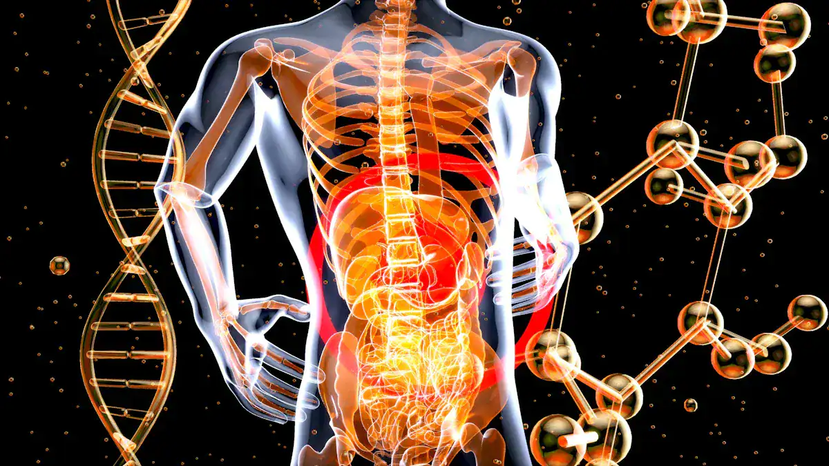
The human body is a truly wondrous and complex machine. Understanding its internal map empowers individuals. Knowing where your organs are located offers practical benefits, from understanding health information to appreciating basic bodily functions. Have you ever wondered exactly where your heart beats or your stomach digests food within your body? Many people lack this basic knowledge of human anatomy.
Knowledge of the location of body organs was generally poor across different human groups, and it has not shown significant improvement over the past three decades.

This highlights a common gap in human understanding of their own body. This guide offers a straightforward approach to demystify organ placement. It provides a quick overview of human anatomy, helping you locate major organs with a clear body organs location chart.
Key Takeaways
Knowing where your organs are helps you understand your body and health better.
Your body has main spaces called cavities that protect important organs like your brain, heart, and lungs.
The thoracic cavity in your chest holds your heart and lungs, while your belly area (abdominal cavity) contains organs like your stomach and liver.
Five vital organs keep you alive: the brain, heart, lungs, kidneys, and liver. Each has a special place and job.
Different organ systems, like the digestive and circulatory systems, work together to keep your body functioning.
Essential Human Anatomy Overview

What Are Organs
Organs are specialized structures within the human body. Each organ performs specific jobs essential for life. For example, the heart pumps blood throughout the body. The lungs oxygenate blood by taking in oxygen and releasing carbon dioxide. The liver converts nutrients, filters blood, and produces bile. Kidneys filter blood and remove waste.
The pancreas produces enzymes for digestion and hormones like insulin. The brain acts as the body’s control center, processing thoughts and sensations. These vital components work together within various organ systems. Understanding the function of each organ is key to understanding human anatomy.
Body Cavities and Regions
The human body contains several main cavities. These cavities are spaces that house and protect organs. Understanding these cavities helps in comprehending human anatomy. This knowledge is fundamental to human anatomy.
The dorsal cavity lies at the back of the body. It includes two parts. The cranial cavity houses the brain, protected by the skull bones. The spinal cavity, also called the vertebral cavity, encloses the spinal cord. The vertebral column protects the spinal cord. This cavity is continuous with the cranial cavity.
The ventral cavity is at the front of the body. It has two main subdivisions. The thoracic cavity is the upper part, enclosed by the rib cage. It contains the lungs and the heart. The diaphragm forms its floor, separating it from the abdominopelvic cavity below it.
The abdominopelvic cavity is the largest cavity. It sits inferior to the thoracic cavity. This cavity is not physically divided by a membrane. It includes the abdominal cavity and the pelvic cavity. The abdominal cavity houses digestive organs. The pelvic cavity contains reproductive organs, some urinary organs, and parts of the digestive system. The abdominal cavity is bounded cranially by the diaphragm. The pelvic cavity is bounded cranially by the abdominal cavity and dorsally by the sacrum. This organization protects vital organs and allows them to function correctly within the human body. The nervous system, with the brain and spinal cord, resides safely within the dorsal cavity. This intricate anatomy ensures the proper functioning of all body systems.
Major Body Cavities and Organ Placement
The human body organizes its internal structures into distinct cavities. These cavities provide protection and specific environments for the organs they contain. Understanding these major body cavities helps us map the precise location of each organ. This knowledge is fundamental to understanding human anatomy.
Thoracic Cavity Organs
The thoracic cavity, located in the chest, protects several vital organs. This cavity houses the heart, lungs, esophagus, thymus, and trachea. It also contains major blood vessels. These vessels transport blood to and from the heart and lungs. They also distribute blood throughout the body and collect it for return to the heart.
The heart, a vital organ, sits centrally within the thoracic cavity. It positions itself between the lungs in the mediastinum. A fibrous membrane sac, the pericardium, encases the heart. This sac separates the heart from other mediastinal structures. The heart’s dorsal surface lies near the vertebrae.
Its anterior surface rests deep to the sternum and costal cartilages. The superior aspect, called the base, is where the great veins and arteries attach. This base is at the level of the third costal cartilage. The inferior tip, or apex, positions slightly to the left of the sternum, between the fourth and fifth ribs. The right side of the heart deflects anteriorly. The left side deflects posteriorly. A depression in the medial surface of the left lung’s inferior lobe, the cardiac notch, accommodates this slight deviation of the apex.
The lungs are paired, cone-shaped organs of respiration. They reside within the thoracic cavity. The heart and other mediastinal structures separate them. Each lung has a base that rests on the diaphragm. An apex extends superiorly approximately 2.5 cm above the clavicle. The lungs possess a medial surface and three borders: anterior, posterior, and inferior. The broad costal surface presses against the rib cage. The smaller mediastinal surface faces medially.
A slit on the mediastinal surface, the hilum, allows entry of the bronchus, blood vessels, lymphatic vessels, and nerves. This forms the lung’s root. The right lung is larger and heavier than the left. The heart’s tilt to the left makes the left lung smaller. It features a cardiac impression to accommodate the heart. This shapes the inferior and anterior parts of its superior lobe into a thin, tongue-like process called the lingula. The human body carefully places these organs for optimal function.
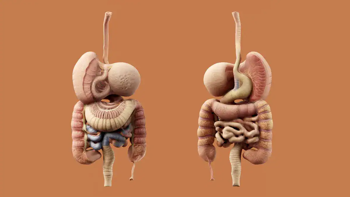
Abdominal Cavity Organs
The abdominal cavity contains the majority of the digestive tract. It also houses the liver, pancreas, spleen, kidneys, and adrenal glands. Many important organs perform crucial functions here.
Here is a list of key organs in the abdominal cavity:
Stomach
Liver
Gallbladder
Spleen
Pancreas
Small intestine
Kidneys
Large intestine
Adrenal glands
Duodenum
Right kidney
Right adrenal gland
Left adrenal gland
The liver, the body’s largest solid organ, primarily sits in the right upper quadrant. It extends into the left hypochondrium. The liver lies inferior to the diaphragm. It reaches as high as the 5th rib on the right side. This vital organ detoxifies the body, regulates glycogen storage, and secretes bile for digestion.
The stomach’s lesser curvature connects to the visceral surface of the liver. The lesser omentum forms this connection. The duodenum is the proximal segment of the small intestine.
It is C-shaped and has four parts: superior, descending, transverse, and ascending. The duodenojejunal junction, where the duodenum connects to the jejunum, is in the left upper quadrant. The jejunum and ileum, other parts of the small intestine, connect to the posterior abdominal wall by the mesentery.
The pancreas, a glandular organ, sits behind the stomach. It secretes insulin and glucagon. It also produces enzymes for digestion and nutrient absorption. The gallbladder, a small hollow sac, sits under the liver. It stores bile the liver produces. The kidneys, two bean-shaped organs, filter blood for waste products. They regulate fluid balance and play a role in blood pressure. The adrenal glands sit atop the kidneys.
Pelvic Cavity Organs
The pelvic cavity is the lower part of the abdominopelvic cavity. It contains organs related to reproduction, excretion, and digestion. This cavity protects several important organs.
Key organs found in the pelvic cavity include:
Reproductive organs
Urinary bladder
Distal ureters
Proximal urethra
Terminal sigmoid colon
Rectum
Anal canal
For females: Uterus, fallopian tubes, ovaries, and upper vagina
Pelvic colon
Bladder
Some sex organs
The urinary bladder’s superior and upper posterior parts are covered by peritoneum. This forms the rectovesical pouch at their junction. In females, the uterus and vagina sit between the urinary bladder and the rectum. The peritoneum covers these structures.
It creates the rectouterine pouch of Douglas between the rectum and the uterus. The vesicouterine pouch is between the posterior surface of the urinary bladder and the anterior surface of the uterus.
The uterus typically anteverts. This places its anterior surface close to the posterosuperior surface of the bladder. The human anatomy ensures precise placement for these delicate organs. The brain, though distant, coordinates the functions of all these body parts.
Understanding the Body Organs Location Chart
A visual body organs location chart helps people understand the complex internal structure of the human body. It shows where each organ sits. This knowledge is fundamental to understanding anatomy. It helps people grasp how different parts of the body work together.
Vital Organ Locations
Five vital organs are essential for life. These include the brain, heart, lungs, kidneys, and liver. Each has a precise place within the body.
The brain sits within the cranial cavity. This cavity is part of the dorsal cavity. The skull bones protect the brain. The heart is situated in the pericardial cavity. This cavity is a component of the thoracic cavity. The lungs are found within the pleural cavities. These cavities flank the pericardial cavity. All these structures reside within the thoracic cavity.
The liver’s primary mass is located in the upper right side of the abdomen. It positions just beneath the rib cage. The kidneys are a pair of organs. They sit on either side of the back. The lower part of the rib cage protects them.
These vital organs also have specific sizes and weights. This table shows their approximate measurements in a human adult.
Organ | Weight (approx.) | Dimensions (approx.) |
|---|---|---|
Brain | 3 pounds (1.36 kg) | Width: 5.5 inches (14 cm), Length: 6.5 inches (16.7 cm), Height: 3.6 inches (9.3 cm) |
Lungs (together) | 2.2 pounds (1 kg) | Height: 9.4 inches (24 cm) |
Heart | Not specified | Length: 4.7 inches (12 cm), Width: 3.3 inches (8.5 cm) |
Liver | Not specified | Not specified |
Kidneys | Not specified | Not specified |
This detailed anatomy helps us appreciate the intricate design of the human form.
Important Organ Placement
Other important organs also have specific placements. These include the gallbladder, pancreas, and stomach. They play crucial roles in digestion and other bodily functions.
The gallbladder is a small, pear-shaped sac. It measures about 3 inches long. It sits on the posterior border of the liver. The cystic duct connects it to the bile ducts of the liver. The pancreas is a gland about 6 inches long. It sits inferior to the stomach.
The duodenum surrounds its medial end. It extends laterally towards the left side of the abdominal cavity. The stomach is a hollow muscular organ. It is about the size of two closed fists. It sits inferior to the diaphragm. It is lateral to the liver on the left side of the abdominal cavity. It forms part of the gastrointestinal tract. It connects the esophagus and the duodenum. This precise anatomy ensures proper digestive processes.
Organ Relationships and Proximity
Organs do not function in isolation. They have close relationships and proximity to each other. This arrangement allows for efficient system operation. A body organs location chart clearly illustrates these connections.
The gallbladder sits next to the liver in the upper right abdomen. The pancreas is located in the upper center to the right portion of the abdomen. It is long and narrow. It works closely with the gallbladder and liver. The stomach’s position between the esophagus and duodenum shows its role in the digestive pathway. The brain coordinates all these functions.
Key Organ Systems and Their Functions
The human body contains many complex systems. Organs work together within these major systems to perform life-sustaining functions. Human anatomy involves 12 major systems. This guide focuses on key systems to detail organ placement and function. Understanding these systems helps comprehend the body’s intricate design.
Digestive System Organs
The digestive system breaks down food and absorbs nutrients. This system starts in the mouth and ends at the anus. The human digestive tract is approximately 23 feet long on average. The small and large intestines together measure about 15 feet or more.
Organ | Function |
|---|---|
Oral Cavity | Ingestion of food, mechanical digestion (chewing), chemical digestion (salivary amylase for carbohydrates, lingual lipase for fats), formation of bolus, swallowing. |
Pharynx | Passageway for food and air, propulsion of bolus to esophagus. |
Esophagus | Propels food from pharynx to stomach via peristalsis. |
Stomach | Mechanical digestion (churning), chemical digestion (pepsin for proteins, gastric lipase for fats), temporary storage of food, production of chyme, absorption of some substances (e.g., alcohol, aspirin). |
Small Intestine | Primary site of chemical digestion (carbohydrates, proteins, fats, nucleic acids) and nutrient absorption, propulsion of chyme. |
Large Intestine | Absorption of water and electrolytes, formation and storage of feces, defecation. |
Liver | Produces bile (for fat emulsification), metabolizes nutrients, detoxifies substances, stores glycogen, produces plasma proteins. |
Gallbladder | Stores and concentrates bile produced by the liver. |
Pancreas | Produces digestive enzymes (amylase, lipase, proteases) and bicarbonate (to neutralize stomach acid), produces hormones (insulin, glucagon) for blood sugar regulation. |
Salivary Glands | Produce saliva (lubrication, salivary amylase, lingual lipase). |
Appendix | Its exact function is unknown, but it is thought to play a role in the immune systems. |
Rectum | Stores feces before defecation. |
Anus | Controls the expulsion of feces from the body. |
Respiratory System Organs
The respiratory system facilitates gas exchange. It takes in oxygen and expels carbon dioxide. The lungs are the primary organs of this system. An adult human male’s average total lung capacity is approximately 6 liters of air. Healthy adult lungs can hold a maximum of about 6 liters of air.
Nasal Cavity: Warms and humidifies inhaled air, traps larger foreign particles with hairs, and contains chemoreceptors for smell and taste.
Pharynx: A tube connects the nasal cavity and mouth to the larynx. It serves as a passage for both air and food.
Larynx (Voice Box): Connects the pharynx and trachea, conducts air, contains vocal cords for sound production, and protects the trachea from aspirated food via the epiglottis.
Trachea (Windpipe): The widest passageway connects the larynx to the lungs for air passage. It branches into two bronchial tubes.
Lungs: These organs take in oxygen for the body’s cells and expel carbon dioxide. They are cone-shaped organs made of spongy, pinkish-gray tissue. A membrane (pleura) surrounds them. The right lung has three lobes, and the left has two.
Bronchi (Large Airways): These tubes carry air into the lungs, branching from the trachea. Main-stem bronchi lead to each lung, then divide into smaller bronchi.
Bronchioles (Small Airways): These are even smaller tubes that branch from the bronchi.
Alveoli (Air Sacs): Tiny air sacs at the end of bronchioles facilitate the exchange of oxygen and carbon dioxide.
Circulatory System Organs
The circulatory system transports blood throughout the body. The heart is the main organ of this system. It pumps blood, delivering oxygen and nutrients to cells. It also removes carbon dioxide and other waste products. The heart controls the rhythm and speed of the heart rate. It maintains blood pressure. An adult heart generally pumps around 5 liters of blood per minute when at rest.
Heart: This muscular organ pumps blood throughout your body.
Arteries: These thin, muscular tubes carry oxygenated blood away from your heart to every part of your body.
Veins: These blood vessels return oxygen-depleted blood to your heart.
Capillaries: These blood vessels connect very small arteries and veins. They allow oxygen, carbon dioxide, nutrients, and waste products to pass into and out of cells.
Urinary System Organs
The urinary system filters blood and produces urine. This system removes wastes, controls fluid balance, and maintains electrolyte levels. The kidneys are the most complex and critical part of the urinary system. Human kidneys typically produce about 1 to 2 quarts of urine daily.
Kidneys: These bean-shaped organs filter blood, produce urine, remove wastes, control fluid balance, and maintain electrolyte levels. You have two, located on either side of the back of your abdomen, below the rib cage. They filter approximately 120 to 150 quarts of blood daily.
Ureters: These two thin tubes, each about 9 inches long, carry urine from your kidneys to your bladder.
Bladder: This hollow, muscular, triangular-shaped organ holds urine until you are ready to empty it. It expands as it fills and can typically hold up to 2 cups of urine.
Urethra: This tube carries urine from your bladder out of your body. Two sphincter muscles close off the urethra to retain urine when not urinating. They open to allow urine to pass.
Nervous System Organs
The nervous system controls all bodily functions. It includes muscle movement, sensory perception, and heart rate. This system is vital for human function and understanding human anatomy. The brain is the central control unit. The human brain contains an estimated 62 to 94 billion neurons.
Organ | Function |
|---|---|
Brain | Controls all bodily functions, including muscle movement, sensory perception, and heart rate. |
Spinal Cord | Connects the brain to nerves below the neck, transmitting electrical impulses between them. |
Nerves | Part of the peripheral nervous system, connecting to every organ and tissue in the body. |
Neurons (nerve cells) | Main cells of the nervous system, responsible for generating, transmitting, and interpreting neural impulses. |
This guide provided a clear body organs location chart. It showed the precise placement and basic functions of major organs within human anatomy. This foundational anatomy knowledge is crucial for understanding the human body. It helps appreciate the intricate design of human anatomy.
FAQ
What are the main body cavities?
The human body has several main cavities. These include the dorsal cavity and the ventral cavity. The dorsal cavity contains the cranial and spinal cavities. The ventral cavity includes the thoracic and abdominopelvic cavities. These spaces protect vital organs. Understanding this basic human anatomy is important.
What organs does the thoracic cavity contain?
The thoracic cavity houses several crucial organs. It contains the heart, lungs, esophagus, and trachea. Major blood vessels also reside here. The rib cage protects these organs. This arrangement is a key part of human anatomy.
What are the five vital organs?
The five vital organs are essential for life. They include the brain, heart, lungs, kidneys, and liver. Each organ performs specific, life-sustaining functions. Their precise placement is critical for overall human anatomy and health.
What is the function of the digestive system?
The digestive system breaks down food. It absorbs nutrients from food. This system starts in the mouth and ends at the anus. Organs like the stomach, small intestine, and large intestine work together. This process is a fundamental part of human anatomy.
What is the primary role of the urinary system?
The urinary system filters blood. It produces urine to remove waste. This system also controls fluid balance. It maintains electrolyte levels in the body. The kidneys are the main organs in this system.