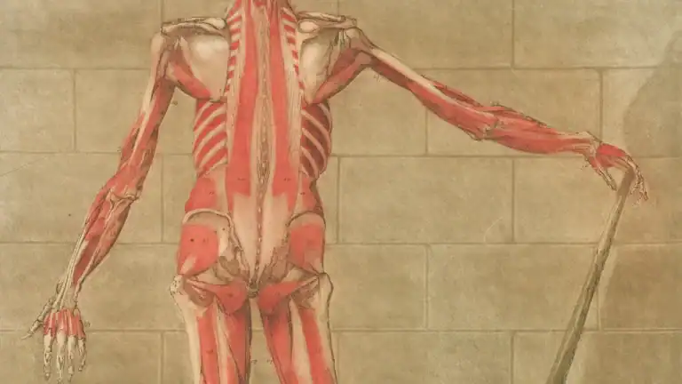
The foot plays a critical role in daily life. It supports the entire body, enables movement, and absorbs shock. This intricate structure boasts 26 bones of the foot, 33 joints, and over 100 muscles, tendons, and ligaments. Approximately 25% of all bones in the human body are in the feet.
This blog provides a comprehensive foot anatomy diagram, mapping the foot’s bones and ligaments. How does this complex foot anatomy allow such mechanics? Tendons connect muscle to bone, while ligaments connect bone to bone.
Key Takeaways
The foot has 26 bones, 33 joints, and over 100 muscles and ligaments. These parts work together for movement and support.
Bones divide into tarsals, metatarsals, and phalanges. The talus and calcaneus are key bones for weight and movement.
Ligaments connect bones and give stability. The plantar fascia supports the arch. Ankle ligaments prevent sprains.
Muscles and tendons move the foot. Extrinsic muscles start in the leg. Intrinsic muscles are inside the foot. They support the arches.
The ankle joint moves the foot up and down. The subtalar joint helps the foot turn in and out. These movements help the foot adapt to different ground.
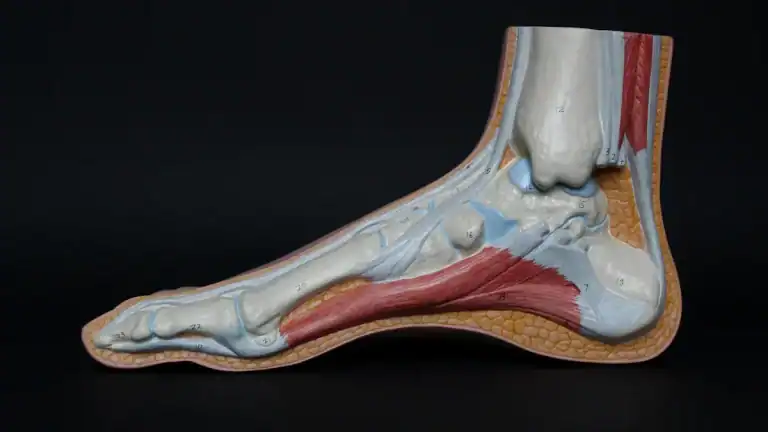
Foot Anatomy Diagram: Bone Structure

The human foot contains 26 bones. These bones divide into three main groups: the tarsals, metatarsals, and phalanges. This foot anatomy diagram helps visualize these structures. These bones of the foot work together, providing support, flexibility, and movement for the entire body.
Tarsal Bones: Hindfoot & Midfoot
The tarsal bones form the hindfoot and midfoot regions. There are seven tarsal bones in each foot. These bones include the talus, calcaneus, navicular, cuboid, and three cuneiform bones (medial, intermediate, and lateral).
Talus (Hindfoot): This bone transmits the body’s weight from the leg to the foot. It also transfers forces from the tibia to the calcaneus.
Calcaneus (Hindfoot): The calcaneus is the largest tarsal bone. It forms the heel and bears the body’s weight when the heel strikes the ground. It also serves as an attachment point for the Achilles tendon.
Navicular (Midfoot): This bone sits on the medial side of the foot. It connects the talus to the cuneiforms and cuboid.
Cuboid (Midfoot): Located on the lateral side, the cuboid sits in front of the calcaneus. It lies behind the fourth and fifth metatarsals.
Cuneiforms (Midfoot): These three wedge-shaped bones (medial, intermediate, lateral) articulate with the navicular and metatarsals. They help form a transverse arch across the foot.

Metatarsal Bones: Midfoot
The midfoot also contains five metatarsal bones. Doctors number them one through five, starting from the big toe side.
These bones connect the tarsal bones to the phalanges. The metatarsal bones are crucial for forming the arches of the foot. These arches are essential for weight-bearing, maintaining balance, and facilitating walking. The metatarsals, with their convex shape, contribute to the foot’s arch. During the late stance phase of gait, the metatarsal acts as a rigid lever for push-off.
Phalanges: Toe Bones
The phalanges are the bones of the toes. Each foot contains 14 phalanges. The great toe (hallux) has two phalanges: a proximal and a distal phalanx. The other four toes each have three phalanges: a proximal, a middle, and a distal phalanx. Occasionally, the little toe may only have two phalanges. The middle and distal phalanges of the fourth and fifth toes can sometimes be fused.
Key Bones: Talus & Calcaneus
The talus and calcaneus are two particularly important bones of the foot. They play critical roles in weight distribution and movement.
The talus has an irregular shape. It divides into three main parts: the head, neck, and body. The head faces forward and carries the articular surface of the navicular bone. The neck is a roughened area between the body and the head. It contains small vascular channels and provides attachment points for ligaments. The body features several prominent articular surfaces.
The trochlea tali on its superior side is semi-cylindrical. It is wider anteriorly than posteriorly. This affects ankle stability based on foot position. No muscles attach to the talus. Its position is determined by neighboring bones. The talus serves as the main bone connecting the lower leg to the foot. It takes on a significant amount of force, especially during twisting or sudden weight application.
The calcaneus is the largest tarsal bone. It bears approximately 50%–60% of body weight in an upright position. The calcaneus has a thin shell of cortical bone. It encloses a sparse but highly organized array of trabecular bone. This cancellous bone, along with high fluid content, allows it to function as a hydrodynamic shock absorber during impact.
The calcaneus provides a lever arm for the Achilles tendon. This enables powerful plantar flexion forces. Its height, width, and infrastructure allow it to withstand high tensile, bending, and compressive forces. A horizontal shelf called the sustentaculum tali arises from its anteromedial portion. It bears the greatest weight per area.
Anatomy of the Foot: Ligament Map

Ligaments are strong, fibrous tissues. They connect bones to other bones. These vital structures provide stability and support throughout the body. In the foot, ligaments create a complex network.
This network holds the 26 bones together. It allows for movement while preventing excessive motion. This section maps out the major ligaments, explaining their location and primary function. Understanding the anatomy of the foot helps one appreciate its complex design.
Plantar Fascia: Arch Support
The plantar fascia is a thick band of connective tissue. It runs along the bottom of the foot. This crucial structure originates in the medial calcaneal tubercle. It ends at the five metatarsal bones. The plantar fascia acts as a crucial tensile element. It prevents the foot from elongating. It also stops the arch from collapsing.
Studies show it contributes about one-quarter of the arch’s stiffness and stability. It also plays a role in the windlass mechanism. Here, the big toe’s dorsiflexion pulls the fascia taut. This action makes the midfoot bones close-packed. It helps support body weight during walking. The plantar fascia is vital for the normal biomechanics of the foot. It provides support for the arch and aids in shock absorption.
Ankle Ligaments: Medial & Lateral
The ankle joint relies on strong ligaments for stability. These ligaments are on both the medial (inner) and lateral (outer) sides of the ankle.
The medial ankle ligament is also known as the deltoid ligament. It is a complex structure. It stabilizes the inner side of the talocrural joint. This ligament has six components. They organize into superficial and deep layers. The deltoid ligament plays a vital role.
It stabilizes the medial malleolus to the talus, calcaneus, and navicular bone. Some components, like the tibiospring ligament, tibionavicular ligament, and deep posterior tibiotalar ligament, are always present. Others, such as the superficial posterior tibiotalar ligament, tibiocalcaneal ligament, and deep anterior tibiotalar ligament, may vary in presence.
On the lateral side, three main ligaments form the lateral collateral ligament complex. These are the anterior talofibular ligament (ATFL), the calcaneofibular ligament (CFL), and the posterior talofibular ligament (PTFL). These ligaments prevent excessive inversion of the foot. They protect the ankle from sprains.
Midfoot Ligaments: Stability
The midfoot region also contains important ligaments. These ligaments ensure stability. The Lisfranc ligament is vital for maintaining foot stability. It connects the metatarsal bones to the tarsal bones. This ligament is the strongest within the Lisfranc joints.
It extends from the lateral side of the medial cuneiform bone. It inserts at the medial side of the base of the second metatarsal bone. Damage to this ligament can lead to instability of the entire midfoot. Strong interosseous ligaments also connect the metatarsal bases. They contribute significantly to the midfoot’s stability.
Other key midfoot ligaments include the plantar calcaneonavicular ligament, also called the spring ligament. This ligament connects bones in the ankle and foot. It provides arch structure. It acts as a passive stabilizer of the plantar arch in the talocalcaneal joint. The calcaneocuboid ligament connects the heel bone to the tarsal bones.
It forms the foot’s arch. This ligament has several parts. These include the medial calcaneocuboid ligament, dorsolateral calcaneocuboid ligament, plantar calcaneocuboid ligament (SPL), and long plantar ligament (LPL). The LPL originates on the inferior surface of the calcaneus. It inserts into the bases of the second to fourth metatarsals. The SPL originates at the anterior tubercle of the calcaneus. It attaches to the plantar surface of the cuboid.
Forefoot Ligaments: Toe Joints
The forefoot also has numerous ligaments. These ligaments stabilize the joints of the toes. They connect the metatarsal bones to the phalanges.
They also connect the individual phalanges to each other. These ligaments are crucial for the fine movements of the toes. They help maintain the foot’s structure during walking and running. They ensure the toe joints remain aligned. This allows for proper push-off during gait.
Foot Anatomy: Joints & Movement
The foot’s complex structure includes many joints. These joints allow the foot to move in various ways. They enable essential functions like walking and running. The ankle (talocrural) joint, subtalar (talocalcaneal) joint, and transverse tarsal joint are primary joints in the foot.
These joints enable motions such as dorsiflexion, plantarflexion, inversion, eversion, pronation, and supination. The metatarsophalangeal joints and interphalangeal joints of the toes also contribute to the foot’s range of motion. They allow for flexion, extension, abduction, and adduction. This section explains how the bones articulate. It focuses on the key joints that enable the foot’s range of motion.
Ankle Joint: Talocrural
The ankle joint, also known as the talocrural joint, connects the lower leg to the foot. This joint is a hinged synovial joint. It primarily allows up-and-down movement. These movements are plantarflexion and dorsiflexion. Plantarflexion points the foot downwards. Dorsiflexion lifts the foot upwards. The talocrural joint is a uniaxial hinge joint. It allows only for these two main movements.
Subtalar Joint: Inversion & Eversion
The subtalar joint sits below the ankle joint. It forms between the talus and calcaneus bones. This joint primarily allows for considerable inversion and eversion movements of the hindfoot. Inversion rotates the sole of the foot towards the midline.
Eversion rotates it laterally. These movements are essential for shock absorption. They also help the foot adapt to uneven ground. The subtalar joint also contributes to supination and pronation. Supination combines inversion, adduction, and plantarflexion. Pronation is the opposite. It results from eversion, abduction, and dorsiflexion.
Midfoot Joints: Tarsometatarsal
The midfoot region contains several important joints. These include the tarsometatarsal joints. They connect the tarsal bones to the metatarsal bones. Other key midfoot joints are the naviculocuneiform joint, cubonavicular joint, and intercuneiform joints. These joints provide stability to the midfoot. They also allow for slight gliding movements. These movements help the foot adapt to different surfaces.
Forefoot Joints: MTP & IP
The forefoot includes the metatarsophalangeal (MTP) joints and interphalangeal (IP) joints. The MTP joints connect the metatarsal bones to the phalanges. They allow the toes to flex, extend, abduct, and adduct. The IP joints are within the toes themselves. They allow the toes to bend and straighten. These joints are crucial for the fine movements of the toes. They help with balance and push-off during walking.
Foot Muscles & Tendons: Movement
Muscles and tendons are vital for the foot’s movement, support, and overall function. Muscles create movement, and tendons connect these muscles to bones. This allows the foot to perform many actions. The foot has two main types of muscles: extrinsic and intrinsic.
Extrinsic Muscles: Leg to Foot
Extrinsic muscles start in the lower leg and extend into the foot. These muscles are primarily responsible for ankle joint motion and balance control. They allow for movements such as eversion, plantarflexion, and dorsiflexion of the foot. For example, the gastrocnemius, soleus, and tibialis posterior muscles are active during running. They help resist foot pronation. The tibialis anterior muscle acts as an invertor during early stance. Peroneus muscles act as evertors. This can increase overall joint stiffness. Other extrinsic muscles include the plantaris, flexor digitorum longus, flexor hallucis longus, and fibularis brevis. The tibialis anterior muscle originates from the tibia. It inserts into the medial cuneiform bone and the base of the first metatarsal bone of the foot.
Intrinsic Muscles: Foot Arches
Intrinsic muscles have both their origin and insertion points within the foot itself. These muscles are crucial for maintaining posture and providing stability to the foot’s arches. Many call them the “core” muscles of the foot. They are similar to how core muscles stabilize the spine. Weakness in these intrinsic muscles can lead to a lower arch height and foot over-pronation. Their main job is to support and stabilize the arches of the foot. They also help maintain the correct position of the big toe. Examples of these muscles include the abductor hallucis, flexor hallucis brevis, abductor digiti minimi, and flexor digitorum brevis.
Major Tendons: Achilles & Tibialis
Two major tendons play critical roles in the foot and ankle. The Achilles tendon, also known as the calcaneal tendon, is at the back of the lower leg. It connects the gastrocnemius and soleus muscles to the calcaneus (heel bone). This is the strongest and thickest tendon in the human body. It enables rising up on toes and pushing forward during walking or running. It also helps stabilize the ankle joint during gait. The tibialis posterior tendon (TPT) originates from the lower leg bones. It courses behind the medial malleolus. It inserts into several bones in the hindfoot and midfoot, including the navicular bone. The TPT is the main dynamic stabilizer of the medial longitudinal arch of the foot. It elevates the medial arch and helps invert and plantar flex the foot.
The foot is a marvel of engineering. This foot anatomy diagram has shown its complex structure. It features 26 bones of the foot, 33 joints, and over 100 muscles, tendons, and ligaments. These bones of the foot, along with other tissues, enable essential functions.
This intricate foot anatomy allows the foot to adapt under load. It spreads out and flattens its arches. This effectively bears weight, propels movement, and absorbs shock. Appreciate the resilience and functional brilliance of the human foot. Understanding its structure helps promote better foot health and injury prevention.
FAQ
What are the main sections of the foot?
The foot divides into three main sections. The hindfoot contains the talus and calcaneus. The midfoot includes the navicular, cuboid, and cuneiforms, along with the metatarsals. The forefoot consists of the phalanges, or toe bones. This structure allows complex movement.
What is the primary role of ligaments in the foot?
Ligaments are strong, fibrous tissues. They connect bones to other bones. In the foot, ligaments provide crucial stability. They hold the 26 bones of the foot together. They also prevent excessive motion while allowing necessary flexibility. The anatomy of the foot relies on these connections.
What movements does the ankle joint allow?
The ankle joint, also known as the talocrural joint, primarily allows two movements. These are plantarflexion and dorsiflexion. Plantarflexion points the foot downwards. Dorsiflexion lifts the foot upwards. This hinge joint is vital for walking and running.
How does the plantar fascia support the foot?
The plantar fascia is a thick band of tissue on the bottom of the foot. It acts as a crucial tensile element. It prevents the foot from elongating. It also stops the arch from collapsing. This structure provides essential arch support and aids in shock absorption.
What is the difference between extrinsic and intrinsic foot muscles?
Extrinsic muscles originate in the lower leg and extend into the foot. They control large movements like ankle plantarflexion. Intrinsic muscles have both their origin and insertion within the foot. They support the foot’s arches and provide fine motor control.