
The human digestive system is a vital process. It converts food into energy and essential nutrients. This complex internal network processes approximately 10 percent of the body’s total energy expenditure daily. Its organs are interconnected, working together seamlessly.
This blog provides a complete digestive system diagram. It identifies each of the parts of the digestive system and explains its role in digestion. Food embarks on a journey through this system, from ingestion to final elimination.
Key Takeaways
The digestive system changes food into energy and nutrients. It uses many organs working together.
Digestion starts in the mouth. It moves through the pharynx, esophagus, stomach, and small and large intestines.
Accessory organs like the liver, gallbladder, pancreas, and salivary glands help digestion. They make important liquids and enzymes.
Food breaks down in stages: ingestion, mechanical digestion, chemical digestion, movement, absorption, and elimination.
Nutrients from food go into the blood. The body uses them for energy, growth, and repair.
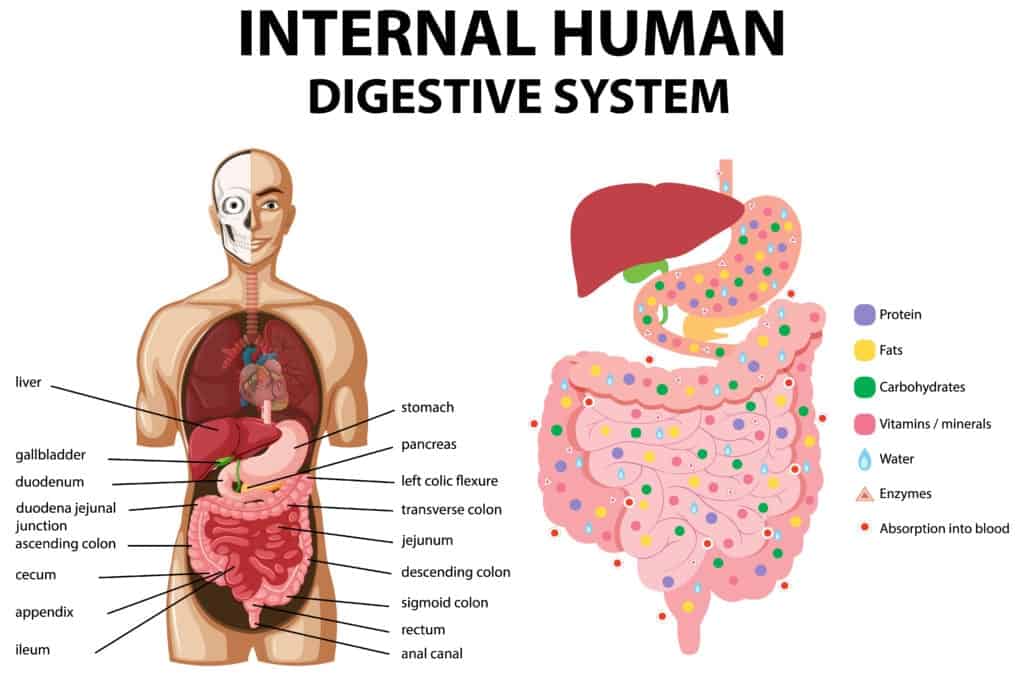
Digestive System Diagram: The GI Tract
The gastrointestinal (GI) tract, also known as the alimentary canal, forms the direct pathway food takes through the body. This long, muscular tube begins at the mouth and ends at the anus. It processes food through various stages, breaking it down and absorbing nutrients. Understanding each of these parts of the digestive system helps clarify the entire process.
The Mouth
Digestion begins in the mouth, also called the oral or buccal cavity. The mouth is framed by the cheeks, tongue, and palate. Lips mark its entrance. Inside, the oral vestibule is a pocket-like area. Gums and teeth border it internally, while cheeks and lips form its external boundaries. The palate forms the roof of the mouth. It acts as a wall between the oral and nasal cavities. The hard palate, located at the front, is bony. It provides a firm surface for the tongue to press food against. The soft palate, mainly muscle, completes the roof of the mouth at the back.
The mouth contains several key structures for initial digestion. Teeth begin mechanical digestion. They grind food into smaller fragments through mastication, or chewing.
The tongue manipulates this chewed food. It forms a soft mass called a bolus, preparing it for swallowing. Salivary glands also play a crucial role. They secrete saliva, which moistens food and cleanses the mouth. Saliva also dissolves food chemicals, allowing for taste.
It contains enzymes that start the chemical digestion of starches. Two important enzymes are present in saliva. Salivary amylase breaks down complex carbohydrates like starch into smaller sugar molecules. Lingual lipase initiates the breakdown of fats into fatty acids and glycerol.
Pharynx and Esophagus
After the mouth, food moves into the pharynx, commonly known as the throat. The pharynx serves as a crucial passageway. It connects the mouth and nose to the esophagus and larynx.
Its main job in digestion is to move swallowed solids and liquids into the esophagus. This process involves the coordinated action of its muscles, especially the pharyngeal constrictors, which are vital for swallowing.
The swallowing process starts voluntarily. The tongue’s strong muscles push the food bolus from the mouth into the pharynx. The soft palate lifts to prevent food from entering the nasal pharynx. The larynx moves upward and forward. Superior pharyngeal constrictor muscles contract, starting a rapid wave-like contraction called peristalsis.
This peristaltic action pushes the bolus down the pharynx. The lower pharyngeal walls elevate to engulf the food mass. The epiglottis diverts the bolus away from the larynx, ensuring it enters the pharynx. The cricopharyngeal muscle, which is the upper esophageal sphincter, relaxes. This allows the bolus to enter the upper esophagus. The pharyngeal peristaltic contraction continues into the esophagus.
Once swallowing begins, the process becomes automatic. The brain signals the muscles of the esophagus. Peristalsis starts, pushing food down the esophagus into the stomach. The muscle behind the food contracts, squeezing it forward. The muscle in front of the food relaxes, allowing movement. These wave-like muscle contractions propel food through the digestive tract.
The Stomach
The stomach is an expansion of the alimentary canal. It is a highly active and muscular organ. It acts as a temporary holding chamber for food. It mixes food with digestive juices and starts the chemical digestion of proteins and triglycerides. The stomach also provides mechanical assistance to digestion. It can hold up to 4 liters of food and fluid.
The stomach has distinct anatomical regions. The cardia is where the esophagus connects to the stomach. Food passes into the stomach here. The fundus is dome-shaped. It sits below the diaphragm, above and to the left of the cardia. The body is the main part of the stomach, located below the fundus. The pylorus is funnel-shaped. It connects the stomach to the duodenum. The pylorus controls stomach emptying into the duodenum through the pyloric sphincter.
Gastric juice, produced in the stomach, contains several key components. Hydrochloric acid (HCl) creates an acidic environment (pH 1.5-3.5). This is necessary for pepsin activation. HCl also denatures proteins, unfolding them for easier enzymatic breakdown.
It kills most bacteria ingested with food, providing a protective barrier. Pepsinogen is an inactive precursor to pepsin, secreted by chief cells. HCl activates it into pepsin. Pepsin is the primary enzyme for protein digestion in the stomach. It breaks down proteins into smaller polypeptides. Gastric lipase has a minor role in fat digestion.
Also It breaks down some triglycerides into fatty acids and monoglycerides, especially in infants. Intrinsic factor is a glycoprotein essential for vitamin B12 absorption in the small intestine.
Mucus forms a protective barrier along the stomach lining. It is rich in bicarbonate, which neutralizes HCl near the stomach wall. This prevents the stomach from digesting itself. Salivary amylase continues to digest carbohydrates in the stomach until the low pH inactivates it. Lingual lipase also continues to digest fats until the low pH inactivates it.
The Small Intestine
The small intestine is a crucial organ for digestion and nutrient absorption. It is where most chemical digestion and absorption of nutrients occur. The small intestine requires a vast surface area for effective nutrient absorption. This extensive surface comes from its long tube and the convolution of its absorptive surface into villi and microvilli.
These structures are crucial for creating enough absorptive surface area for intestinal function. A larger surface area is vital for efficient absorption and exchange of materials, especially for nutrient uptake in the digestive system. The small intestine is specifically adapted for this role. Villi and microvilli significantly increase its surface area, maximizing nutrient absorption.
The small intestine has three segments. The duodenum is the initial and shortest segment. Digestion continues here with the help of bile and pancreatic juices. Preparation for nutrient absorption begins. The jejunum is the middle section. It is primarily responsible for absorbing small nutrient particles that have been previously digested.
The ileum is the final segment. Its main role is to absorb vitamin B12, bile salts, and any remaining digestive products not absorbed earlier. Nutrient absorption occurs from the intestinal surfaces. The duodenum, jejunum, and ileum have an expansive mucosa due to villi and microvilli on enterocytes. Villi are small, finger-like projections that increase surface area for efficient nutrient absorption. They contain blood vessels and lacteals to transport absorbed nutrients.
Microvilli cover the villi. They contain enzymes that further break down nutrients, enhancing absorption in the small intestine. Refluxive motility and villi movement in the small intestine facilitate contact between lumen contents and the unstirred water layer at the villi surfaces. Contractile elements move microvilli to enhance contact after transfer to the surface. The small intestine is a highly efficient organ.
The Large Intestine
The large intestine follows the small intestine. It plays a vital role in processing waste material. Its primary functions include absorbing electrolytes and water, propelling intestinal contents, forming and temporarily storing feces, and defecation.
The large intestine has several main sections. The cecum is the initial segment. It is involved in digestion and temporary storage of chyme. It also reabsorbs fluid and electrolytes. The vermiform appendix, a blind lymphoid pouch, originates from the cecum. It contributes to maintaining gut flora and mucosal immunity. The ascending colon is the first of the four main sections. It extracts water and other essential nutrients from waste material for recycling. This process begins after waste exits the small intestine via the ileocecal valve and passes through the cecum.
Peristalsis moves the waste upwards towards the transverse colon. The transverse colon spans between the right and left colic flexures. The descending colon extends from the splenic flexure to the start of the sigmoid colon. Its role is to store feces before they are emptied into the rectum. The sigmoid colon, rectum, and anal canal complete the large intestine.
The gut microbiota in the large intestine performs several important functions. They ferment non-digestible substrates like dietary fibers and intestinal mucus. This supports the growth of specialized microbes. They also contribute to bile acid metabolism, producing unconjugated and secondary bile acids. These act as signaling molecules and metabolic regulators.
The microbiota produce short-chain fatty acids (SCFAs). Acetate is the most abundant SCFA. The body uses it in cholesterol metabolism and lipogenesis. Propionate transfers to the liver. It regulates gluconeogenesis and satiety signaling. Butyrate is the main energy source for human colonocytes. It can induce apoptosis of colon cancer cells. It also activates intestinal gluconeogenesis, which benefits glucose and energy homeostasis. Butyrate maintains oxygen balance in the gut to prevent dysbiosis.
Rectum and Anus
The rectum is the terminal segment of the large intestine. It connects the colon to the anus. It is approximately 12-15 cm long. It features sacral and anorectal flexures, along with three lateral flexures known as Houston valves. These help support the weight of feces and prevent urgent defecation.
The rectal ampulla, located at the end of the rectum, serves as a temporary holding area for excrement before release. The rectum acts as a storage site for waste before elimination through the anus. It absorbs water and electrolytes, thickening feces.
When feces enter the rectal ampulla, stretch receptors in the rectal wall activate. They signal the need to defecate to the central nervous system. The rectum contributes to defecation by contracting. Along with voluntary tension of the diaphragm and abdominal muscles, this increases intra-abdominal pressure.
The anus is the final opening of the digestive tract. It is controlled by two sphincters: the internal and external anal sphincters. When the rectum is full, nerves trigger the urge for a bowel movement.
The internal anal sphincter automatically relaxes. This sphincter is formed by the circular muscle layer of the rectum. It is responsible for the resting tone of the anus. The external anal sphincter is under conscious control. This allows activation when an individual is ready for defecation. It surrounds the internal sphincter and extends to the anal verge.
It is a fatigable muscle composed of both ‘slow twitch’ and ‘fast twitch’ fibers. It is primarily responsible for voluntary control rather than resting tone. Both sphincters work individually and together in response to rectal distension and the sensation of rectal filling.
Rectal sensory awareness at a critical filling level triggers the perception of the need to evacuate. In a socially appropriate setting, adopting a sitting or squatting position straightens the rectal angle. Rectal contents provoke reflex relaxation of the anal sphincters and puborectalis. A Valsalva maneuver is performed, raising abdominal pressure and tensing anterior abdominal wall muscles. Pelvic floor relaxation allows stool to enter the lower rectum, initiating spontaneous recto-sigmoid contractions.
Large propulsive contractions of the rectum continue until it is empty, maintained by sensory input from the anus. As stool passes, it stretches the external anal sphincter. After the last bolus, a ‘closing reflex’ stimulates to maintain continence. This complex coordination ensures efficient waste elimination from the digestive system diagram.
Key Parts of the Digestive System: Accessory Organs
Accessory organs play a crucial role in digestion. They aid the process without directly contacting food. These organs produce and secrete essential substances. These substances help break down food and absorb nutrients. Understanding these parts of the digestive system is key to grasping the entire digestive process.
Salivary Glands
Salivary glands produce saliva. Saliva is mostly water, about 99.5%. It also contains electrolytes, mucus, glycoproteins, enzymes, and antibacterial compounds. These components give saliva many important functions.
Saliva keeps the mouth and throat moist and comfortable. It protects the delicate lining from sticking and harmful substances. Saliva moistens food, making it easier to chew and swallow. This helps form a soft mass called a bolus. The enzyme amylase in saliva starts the digestion of starches.
Lysozyme, another component, protects against infection by breaking down bacteria. Saliva also protects teeth. It contains calcium hydroxyapatite, which prevents demineralization. It washes away food debris and dilutes acidic foods. This reduces cavities and gum disease.
Saliva helps maintain a pH balance of 6.0 to 7.5 in the mouth. Proteins and growth factors in saliva help repair tissue in the mouth, promoting wound healing. Saliva also helps taste food. It provides moisture for taste buds to detect flavors.
Different types of salivary glands produce different secretions:
Serous secretions primarily contain alpha-amylase. This enzyme breaks down starch into maltose and glucose.
Mucous secretions primarily contain mucin. Mucin acts as a lubricant.
The body has several major salivary glands:
Salivary Gland Type | Primary Secretion Type |
|---|---|
Parotid gland | Purely serous saliva |
Submandibular gland | Both serous and mucous (3:2 ratio) |
Sublingual gland | Mainly mucous saliva |
Minor salivary glands | Mainly mucous saliva |
These glands work together. They ensure the mouth has the right environment for initial digestion and protection.
The Liver
The liver is the largest internal organ. It performs many vital functions. In digestion, the liver’s main role is producing bile. Bile is a digestive fluid. The liver continuously produces bile. It then sends bile to the gallbladder for storage and concentration.
Bile primarily aids in the digestion of lipids, or fats. It does this through a process called emulsification. Emulsification breaks down large lipid globules into smaller ones. This distributes them widely in the chyme, which is the partially digested food from the stomach.
Bile salts are key to this process. They have both hydrophobic (water-fearing) and hydrophilic (water-loving) sides. This allows them to interact with both lipids and water. They surround lipid droplets, forming micelles. The hydrophobic sides face the fat, and the hydrophilic sides face outwards.
These outward-facing sides are negatively charged. This prevents fat droplets from clumping back together.
Emulsification is crucial because lipases, the enzymes that digest fats, can only efficiently act on lipids when they are broken into small aggregates. This dispersion into micelles significantly increases the surface area for pancreatic lipase to work.
Lipases then break down lipids into fatty acids and glycerides. These smaller molecules can then pass into the intestinal lining cells. Bile also enhances the absorption of fat-soluble vitamins (A, D, E, K).
Without bile salts, most dietary lipids would pass through the body undigested. This leads to a condition called steatorrhea. Steatorrhea can cause deficiencies in essential fatty acids and fat-soluble vitamins.
The Gallbladder
The gallbladder is a small, pear-shaped organ. It sits just under the liver. Its main purpose is to store and concentrate bile produced by the liver. When bile enters the gallbladder, it undergoes a concentration process.
The gallbladder removes water and electrolytes, especially sodium and chloride ions, from the bile. This process can concentrate bile three to ten times its original strength. This significantly enhances its digestive capabilities. The concentrated bile then waits until the body needs it.
When a person eats food containing fats, the gallbladder receives signals to contract. These signals come from hormones and nerves. The gallbladder squeezes the stored bile through the biliary tract.
Bile travels down the cystic duct. It then goes directly into the duodenum, the first section of the small intestine. There, it mixes with the food contents. The sphincter of Oddi, a muscular valve, relaxes and opens at the same time. This allows bile to flow from the gallbladder into the small intestine to aid digestion.
The Pancreas
The pancreas is a gland located behind the stomach. It has two main functions: producing digestive enzymes and regulating blood sugar.
The pancreas produces a variety of key digestive enzymes. These enzymes are crucial for breaking down carbohydrates, proteins, and fats in the small intestine.
Pancreatic amylase breaks down starch and glycogen.
Pancreatic lipase degrades triglycerides into two fatty acids and a monoglyceride.
Trypsinogen is an inactive protease. It activates to trypsin in the duodenum. Trypsin breaks down proteins at basic amino acids.
Chymotrypsinogen is another inactive protease. It activates to chymotrypsin. Chymotrypsin breaks down proteins at aromatic amino acids.
Carboxypeptidase removes terminal amino acid groups from proteins.
Elastases degrade elastin and other proteins.
Nucleases (DNAase, RNAase) degrade nucleic acids.
Other enzymes include sterol esterase and phospholipase.
These pancreatic enzymes further process proteins into smaller peptides and amino acids. Pancreatic lipase specifically hydrolyzes a triglyceride molecule into two fatty acid molecules and a monoglyceride.
The pancreas also plays a vital role in regulating blood sugar levels. Blood glucose, or blood sugar, is the primary sugar in the blood. It is the body’s main energy source, coming from food. When blood glucose levels increase, it signals the pancreas to release insulin. Insulin is a hormone.
It helps glucose enter cells for energy use. Insulin also tells the liver to store excess blood sugar for future energy needs. This process helps blood sugar levels return to normal.
The Human Digestive System: Journey and Processes
The human body performs a complex series of actions to get nutrients from food. All organs work together in a coordinated way. This entire process of digestion involves several distinct stages.
Digestion Stages
The body breaks down food and absorbs nutrients through a multi-step process. This process of digestion begins when food enters the mouth.
Ingestion: Food enters the mouth.
Mechanical Digestion: Physical actions break down large food pieces. Chewing in the mouth and churning in the stomach are examples.
Chemical Digestion: Enzymes transform complex food molecules into simpler ones. This happens through hydrolysis.
Movements: Food moves through the digestive tract. Swallowing, mixing in the stomach, and peristalsis propel food.
Absorption: Simple molecules pass into the blood or lymph capillaries. This happens through the small intestine lining.
Elimination: The body removes undigested materials.
Mechanical digestion breaks food into smaller particles. These physical processes alone cannot reduce food to absorbable micronutrients. Chemical digestion then uses enzymes. These enzymes further break down particles into their smallest components. The small intestine’s continuous movements expose food particles to digestive enzymes. This enhances chemical digestion. It also moves food towards the large intestine.
Nutrient Absorption
After digestion, the body absorbs nutrients. This is a vital part of the process of digestion. The small intestine is the main site for this.
Carbohydrate Absorption: The body absorbs all carbohydrates as monosaccharides (glucose, galactose, fructose). Glucose and galactose enter cells with sodium ions. Fructose enters cells by facilitated diffusion.
Protein Absorption: Most proteins absorb as amino acids. This happens mainly in the duodenum and jejunum. Amino acids enter cells through active transport, often with sodium. Short chains of dipeptides and tripeptides also enter cells actively. They break down into amino acids before entering the blood.
Fat Absorption: Short-chain fatty acids absorb by simple diffusion into cells and then enter the blood. Long-chain fatty acids and monoacylglycerides form micelles with bile salts. They then diffuse into cells. Inside cells, they become triglycerides. These form chylomicrons, which enter lymphatic vessels.
The gastrointestinal tract also absorbs vitamins, minerals, and water. Fat-soluble vitamins (A, D, E, K) need bile salts for absorption. Water-soluble vitamins absorb directly into the bloodstream. Water absorption occurs throughout the GI tract. Osmotic gradients drive this process. Minerals also absorb across the digestive tract.
Waste Elimination
The final stage of the digestive system diagram involves waste elimination. The body removes undigested materials. Feces are the waste product.
Composition of Feces:
75% water and 25% solid matter.
Solid matter includes dead bacteria (30%), indigestible food (30%), fats (10-20%), inorganic substances (10-20%), and protein (2-3%).
Other components include cell debris, bile pigments, and dead leukocytes.
Bacterial action influences the brown color and odor of feces. The gastrointestinal tract eliminates nitrogenous waste as feces. It also removes salts, glucose, calcium, iron, and fiber. This completes the journey through the digestive system.
The human digestive system is a marvel of coordination. Each organ plays a vital role, from the mouth’s initial breakdown to the large intestine’s waste processing. A healthy digestive system is crucial for overall well-being. It ensures:
Regular and pain-free bowel movements
Consistent energy levels
Normal amounts of gas and bloating
Mental clarity and focus
Understanding these intricate processes helps us appreciate our body’s internal workings. It also encourages further learning about digestive health. The body’s efficiency and biological marvel are truly astounding.
