The knee joint plays a critical role in daily activities and mobility. This complex joint allows us to walk, run, and jump. Understanding each bone, ligament, and muscle provides insight into its basic knee anatomy. This knowledge helps maintain overall health and prevents injury. Studies show nearly 25% of American adults experience chronic knee pain annually.
This condition also places a significant economic burden on healthcare systems. This guide explores the essential bones, ligaments, and muscles of the knee, offering a comprehensive look at knee joint anatomy.
Key Takeaways
The knee joint is complex. It helps you walk, run, and jump. It has bones, ligaments, and muscles.
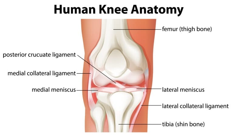
The main bones are the femur (thigh bone), tibia (shin bone), and patella (kneecap). Ligaments connect these bones and keep the knee stable.
Cartilage and menisci are like cushions. They help the knee move smoothly and absorb shock. This protects the knee from damage.
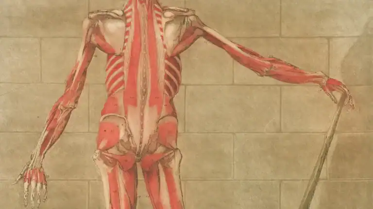
Muscles like the quadriceps and hamstrings move the knee. Tendons connect these muscles to the bones. They help you bend and straighten your leg.
Taking care of your knees is important. You can do this by strengthening muscles and stretching. This helps prevent injuries and pain.
Knee Joint Anatomy Overview
Defining the Knee Joint
The knee joint is a remarkable structure. It functions as a synovial joint, allowing for smooth movement. It also acts as a complex hinge joint, primarily facilitating flexion and extension. This intricate design allows for a wide range of motion. The knee joint anatomy involves two main articulations.
The tibiofemoral articulation connects the medial and lateral condyles of the femur with the tibial condyles. This part of the joint bears the body’s weight. The patellofemoral articulation involves the anterior aspect of the distal femur and the patella, or kneecap.
This articulation improves the efficiency of the quadriceps femoris muscle. It allows the quadriceps tendon to insert directly over the knee. Understanding this complex joint helps in appreciating its function.
Key Components
Several essential components make up the knee. Each part plays a vital role in the overall function of the knee. The primary bones involved are the femur, tibia, and patella. Strong bands of tissue, called ligaments, connect these bones. Ligaments provide stability to the joint. Muscles surround the knee, enabling movement. Tendons connect these muscles to the bones. Cartilage and menisci also contribute to the knee’s structure.
Articular cartilage covers the ends of the bones, ensuring smooth gliding. The menisci act as cushions, absorbing shock and further stabilizing the knee joint. This integrated system allows the knee to perform its critical functions in daily activities.
Bones of the Knee Joint
The knee joint relies on several key bones for its structure and function. These bones work together to support body weight and allow movement. Understanding each bone helps clarify the overall knee anatomy.
Femur: Thigh Bone
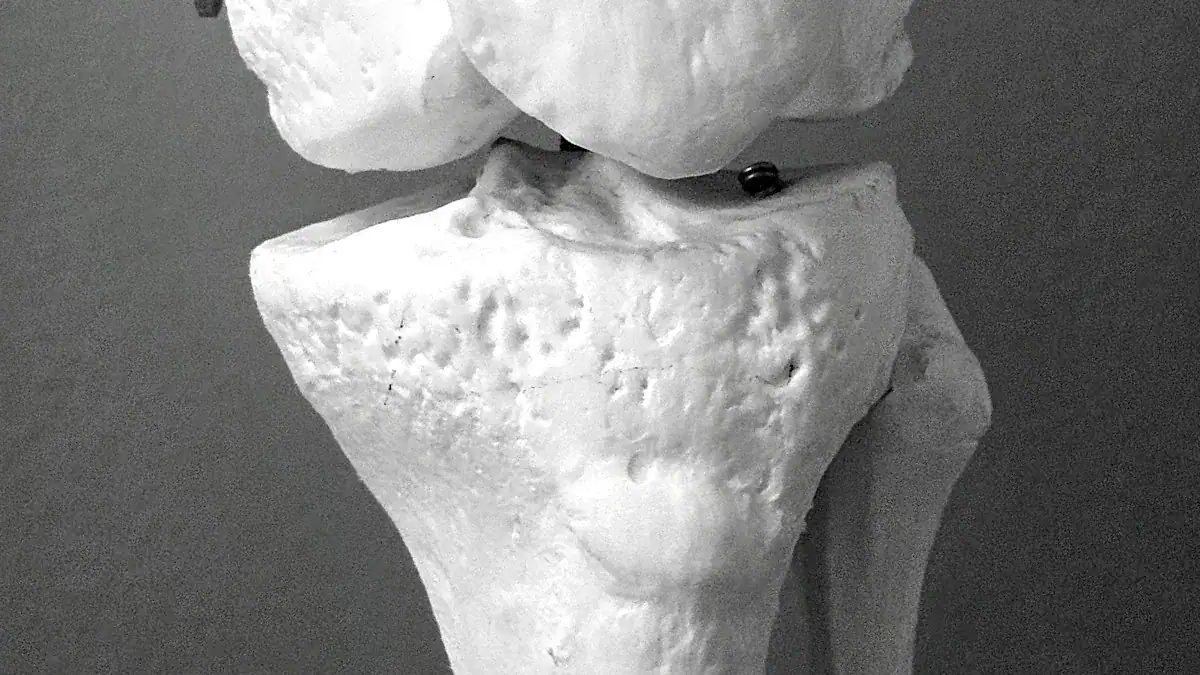
The femur is the longest and strongest bone in the human body. It forms the upper part of the knee joint. The femur’s lower end has two rounded projections, called condyles. These condyles articulate with the tibia. The femur plays several critical roles in the knee’s biomechanics:
It transmits body weight to the tibia.
It handles significant forces; the tibiofemoral joint reaction force can be three times body weight during walking and four times during climbing.
Its interaction with the tibia allows knee motion in the sagittal plane, from 3 degrees of hyperextension to 155 degrees of flexion.
It helps with knee rotation, including the posterior rollback mechanism during flexion.
It provides attachment points for crucial ligaments like the anterior cruciate ligament (ACL) and posterior cruciate ligament (PCL). These ligaments prevent the knee from moving too far forward or backward.
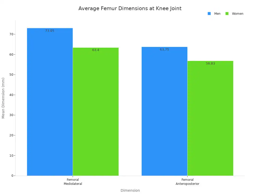
The femur also articulates with the patella, forming the patellofemoral joint. This complex allows for efficient locomotion and helps absorb and redistribute forces. Researchers have measured the average dimensions of the adult femur at the knee joint. These measurements show differences between men and women.
Dimension | Men (Mean ± SD) | Women (Mean ± SD) |
|---|---|---|
Femoral Mediolateral (FML) | 73.05 ± 4.01 mm | 63.40 ± 3.32 mm |
Femoral Anteroposterior (FAP) | 63.75 ± 3.70 mm | 56.83 ± 3.37 mm |
The femoral mediolateral dimension measures the width between the outer aspects of the medial and lateral femoral condyles. The anteroposterior dimension measures the widest distance from the front to the back of the femoral condyles.
Tibia: Shin Bone
The tibia is the larger of the two lower leg bones. It forms the lower part of the knee joint. The tibia bears most of the body’s weight. Its top surface, called the tibial plateau, has two condyles that articulate with the femur. The tibia has specific features that contribute to knee stability.
The intercondylar eminence sits between the tibial condyles. It projects upwards as medial and lateral intercondylar tubercles. This area serves as the main attachment site for the knee joint’s ligaments and menisci. These structures are vital for keeping the knee stable. The tibial condyles connect with the femoral condyles, forming the main knee joint and ensuring its structural strength.
Patella: Kneecap
The patella, or kneecap, is a small, triangular bone located at the front of the knee. It sits within the quadriceps tendon. The patella is a sesamoid bone, meaning it is embedded within a tendon. This unique position gives the quadriceps muscles a significant mechanical advantage.
The patella moves the quadriceps tendon further away from the knee’s rotation point. This increased distance means less force is needed for knee extension. The patella makes the quadriceps approximately 30% more powerful. This biomechanical advantage is especially clear during full knee extension. People who have had their patella removed often find it very hard to fully extend their knees. They lose the mechanical advantage the patella provides.
The patella’s contribution to mechanical advantage changes with knee flexion. It can increase the lever arm by 30% at 45 degrees of flexion. This mechanism effectively boosts the knee extensor moment from quadriceps contraction.
Fibula’s Role
The fibula is the smaller bone of the lower leg. It runs parallel to the tibia. The fibula does not directly form part of the knee joint. However, it plays an indirect role in knee stability and muscle attachment.
The fibula’s head, or apex, provides attachment for the fibular collateral ligament of the knee joint. A portion of the biceps femoris muscle tendon also attaches here. The fibula primarily functions as an attachment site for many muscles. It also helps brace the tibia against bending and twisting forces.
Popliteofibular ligament: This ligament connects the popliteus muscle to the medial aspect of the fibula. It is on the posterolateral side of the knee.
Fabellofibular ligament: This ligament connects a small bone on the femur to the posterolateral edge of the fibula’s styloid process.
Other muscles like the extensor digitorum longus, peroneus longus, and soleus attach to the fibula. The flexor hallucis longus and peroneus brevis also have attachments.
Knee Ligaments
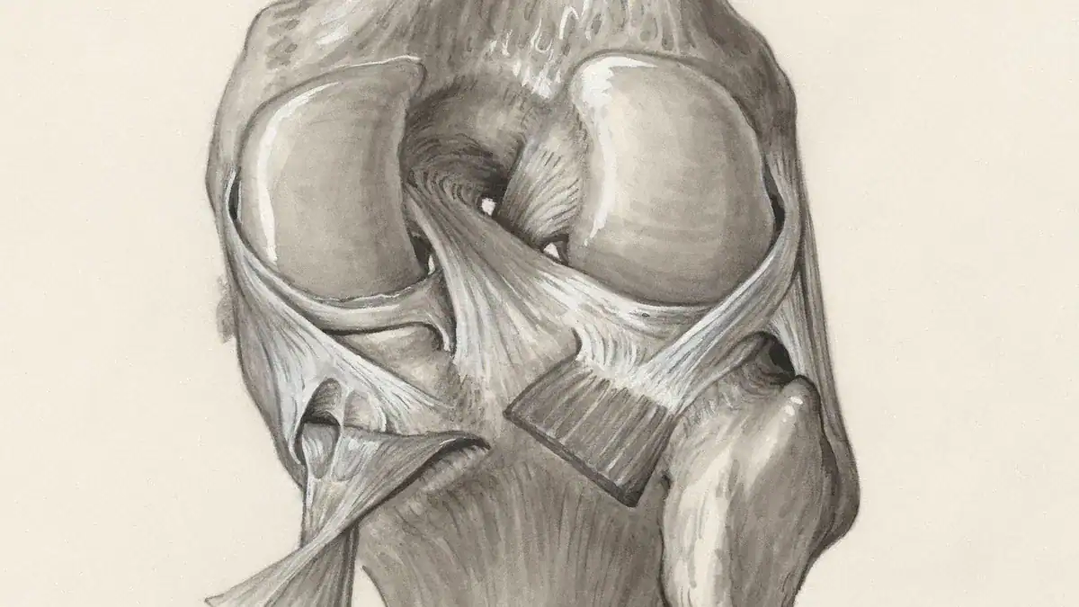
Ligaments are tough bands of tissue. They connect bones. These strong structures are vital for knee stability. They hold the knee joint together. The knee has two main groups of ligaments. These groups provide crucial support.
Cruciate Ligaments (ACL, PCL)
The cruciate ligaments form one group. They cross each other inside the knee joint. These are the anterior cruciate ligament (ACL) and the posterior cruciate ligament (PCL).
The anterior cruciate ligament (ACL) is a key ligament. It originates from the anteromedial aspect of the intercondylar area on the tibial plateau. It extends upwards and backwards. It attaches to the posteromedial aspect of the lateral femoral condyle. The ACL consists of two bundles. An anteromedial bundle (AMB) attaches from the roof of the intercondylar notch. A posterolateral bundle (PLB) attaches from the wall of the intercondylar notch.
Structure | Origin | Insertion |
|---|---|---|
Anterior Cruciate Ligament (ACL) | Anterior intercondylar area of the tibia | Posteromedial side of the lateral femoral condyle |
The ACL prevents the shin bone from sliding out in front of the thigh bone. It also prevents anterior displacement of the tibia. It stops the posterior rolling of the femur during flexion. The posterior cruciate ligament (PCL) is also important.
It prevents the shin bone from sliding backward under the femur. The PCL prevents excessive posterior displacement of the tibia in relation to the femur. These ligaments work together. They control the front-to-back movement of the knee.
Collateral Ligaments (MCL, LCL)
The collateral ligaments form the second group. They are on the sides of the knee. These include the medial collateral ligament (MCL) and the lateral collateral ligament (LCL).
The medial collateral ligament (MCL) is on the inner side of the knee. It has a superficial layer.
This layer attaches proximally just posterior to the medial femoral epicondyle. It attaches distally approximately 6 cm beyond the medial tibial plateau. The MCL’s deep layer is a thickening of the joint capsule. It includes meniscofemoral and meniscotibial components.
The lateral collateral ligament (LCL) is on the outer side of the knee. It originates proximally just posterior to the lateral femoral condyle. It attaches distally to the proximal portion of the fibula. It joins with the biceps femoris tendon there.
The medial collateral ligament (MCL) is the primary restraint to valgus rotation. Valgus rotation means the knee bends inward. Sectioning the MCL leads to significant valgus instability.
In full extension, the MCL resists about 50% of the applied valgus moment. It acts as a secondary medial stabilizer. The LCL helps prevent the knee from bending outward. It resists varus stress. These ligaments provide side-to-side knee stability.
Ligament Function
Ligaments are crucial for knee stability. They connect the bones of the knee joint. This connection limits excessive movement. They guide the knee through its normal range of motion.
The cruciate ligaments control front-to-back movement. The collateral ligaments control side-to-side movement. Together, these ligaments ensure the knee joint remains stable. They protect it from injury during daily activities and sports.
Knee Cartilage and Menisci
The knee joint contains specialized tissues that ensure smooth movement and absorb impact. These tissues are articular cartilage and the menisci. They are vital for the knee’s long-term health and function.
Articular Cartilage
Articular cartilage is a smooth, slippery tissue. It covers the ends of bones within the knee joint. This specialized connective tissue is hyaline cartilage. It allows bones to glide over each other with very little friction. This smooth surface is essential for comfortable movement of the knee.
Articular cartilage has a complex structure. It features a dense extracellular matrix and scattered chondrocytes. The matrix primarily contains collagen, proteoglycans, and glycoproteins. These components help the cartilage retain water molecules.
Water makes up to 80% of its wet weight. This water content significantly influences the cartilage’s mechanical properties. Collagen, mainly type II, provides tensile strength. Proteoglycans, like aggrecan, give compressive strength. They retain water through osmotic pressure, which helps maintain the collagen mesh tension.
Chondrocytes are specialized cells. They produce collagen, proteoglycans, and other matrix proteins. Articular cartilage lacks blood vessels, nerves, and lymphatic tissue. It receives nutrition through synovial fluid diffusion.
This tissue has multiple zones:
Superficial Zone: This outer layer resists shear forces. Its collagen fibers are arranged tangentially.
Transitional (Middle) Zone: This zone resists compression. It has collagen fibers arranged obliquely.
Radial (Deep) Zone: This layer also resists compression. It contains the largest collagen fibrils and the highest proteoglycan concentration.
Tidemark: This zone resists shear and acts as a calcification front.
Calcified Zone: This zone anchors the articular cartilage to the underlying bone.
Menisci: Cushions of the Knee
The menisci are C-shaped pieces of cartilage within the knee. There are two menisci in each knee: the medial meniscus and the lateral meniscus. They sit between the femur and the tibia. These structures act as cushions. They help distribute weight evenly across the knee joint.
The menisci perform several critical functions. Their primary role is tibiofemoral load transmission. They spread the forces across the joint surfaces. This prevents concentrated pressure on one spot. The menisci also contribute to shock absorption.
They absorb impact during both repetitive movements and sudden loads. Studies show that an intact meniscus has the highest shock-absorbing potential. Removing meniscus tissue, even partially, significantly lowers this potential. This confirms the menisci’s vital role in cushioning the knee. A torn meniscus is a common injury that can impair these functions.
Knee Muscles and Tendons
Muscles and tendons work together to move the knee. Muscles create movement. Tendons connect these muscles to bones. This section details the major muscle groups that move the knee. It also explains the tendons that link them to the bones.
Quadriceps Femoris
The quadriceps femoris is a large muscle group. It sits on the front of the thigh. This group consists of four individual muscles. They are the primary movers for knee extension. This means they straighten the leg at the knee.
Muscle Name | Contribution to Knee Extension |
|---|---|
Rectus femoris | The rectus femoris crosses both the hip and knee joints. It extends the knee and also helps flex the hip. |
Vastus lateralis | The vastus lateralis is the largest quadriceps muscle. It sits on the side of the thigh. It is a powerful extensor of the knee. |
Vastus medialis | The vastus medialis sits on the inner side of the thigh. It is important for the last 10-20 degrees of knee extension. It also helps stabilize the patella. |
Vastus intermedius | The vastus intermedius lies deep to the rectus femoris. It sits between the vastus lateralis and vastus medialis. It is a pure knee extensor. |
These muscles work together. They allow actions like kicking, standing up, and climbing stairs.
Hamstrings
The hamstrings are a group of three long muscles. They are located on the back of the thigh. These muscles are the biceps femoris, semimembranosus, and semitendinosus muscles. All hamstring muscles cross both the hip and knee joints. Their main job is to flex the knee joint. They also extend the hip.
The biceps femoris has two heads. Its long head acts on both the hip and knee. It helps with hip extension and knee flexion. The short head of the biceps femoris acts only on the knee joint. It contributes to knee flexion. Both heads together help bend the leg at the knee joint. The semimembranosus and semitendinosus muscles also help bend the knee.
Semimembranosus: This muscle contributes to knee flexion and hip extension.
Semitendinosus: This muscle also contributes to knee flexion and hip extension.
Biceps Femoris long head: This part of the muscle contributes to knee flexion and hip extension.
Calf Muscles
The calf muscles are on the back of the lower leg. They also influence the knee. The medial gastrocnemius (MG) and lateral gastrocnemius (LG) are key calf muscles. They help with plantar flexion, which points the foot downwards.
The stiffness of the MG and LG is much higher when the knee is fully extended. It is lower when the knee is bent to 90 degrees. This shows their effect on knee flexion.
The gastrocnemius (GM) is a biarticular muscle. This means it crosses both the knee and ankle joints. It can influence both knee flexion and ankle plantarflexion.
The soleus (SOL) muscle is another important calf muscle. It helps with plantarflexion. Its stiffness is higher than the MG and LG, regardless of knee position. The soleus is a monoarticular plantar flexor. It mainly acts on the ankle joint for plantarflexion.
Knee Tendons
Tendons are strong, fibrous cords. They connect muscles to bones. They transmit the force from muscle contractions to the bones. This allows movement. Several important tendons are around the knee joint.
Quadriceps Tendon: This tendon connects the quadriceps muscles to the patella. It sits just above the kneecap.
Patellar Tendon: This tendon connects the patella to the tibia. It runs from the bottom of the kneecap to the shin bone.
Hamstring Tendon: These tendons connect the hamstring muscles to the knee joint. They are at the back of the leg.
These tendons are essential for the knee’s function. They allow the powerful leg muscles to move the knee effectively.
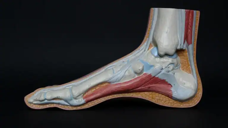
Knee Diagram
Knee Joint Function
Integrated Knee Function
The knee joint performs many actions. It allows people to walk, run, and jump. This complex joint works because all its parts function together. Bones provide the structure. Ligaments connect these bones and give stability. Muscles move the bones. Cartilage and menisci ensure smooth motion and absorb impacts. Each component plays a vital role. The knee joint allows for both strong support and flexible movement. This integrated system makes the knee a powerful and adaptable joint in the human body.
Knee Range of Motion
The knee joint moves in specific ways. It primarily performs flexion and extension. Flexion means bending the knee. Extension means straightening the leg. Most adults achieve a functional range of 0 to 135 degrees. Full extension is 0 degrees, meaning a straight leg. Full flexion can reach 135 to 150 degrees, allowing the heel to touch the buttock.
The typical range of motion varies slightly by age and gender.
Motion | Females (20-44 years) | Males (20-44 years) | Females (45-69 years) | Males (45-69 years) |
|---|---|---|---|---|
Knee flexion | 141.9 (140.9 – 142.9) | 137.7 (136.5 – 138.9) | 137.8 (136.5 – 139.1) | 132.9 (131.6 – 134.2) |
Knee extension | 1.6 (1.1 – 2.1) | 1.0 (0.6 – 1.4) | 1.2 (0.7 – 1.7) | 0.5 (0.1 – 0.9) |
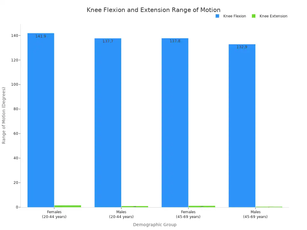
These ranges show how much the knee can bend and straighten. Maintaining a good range of motion helps with daily activities.
Weight Bearing and Shock Absorption
The knee joint carries the body’s weight. It also absorbs shocks from movement. Every step a person takes puts stress on the knee. The menisci and articular cartilage play key roles in this function. Menisci act as cushions. They spread the forces across the joint surfaces. This prevents too much pressure on one spot. Articular cartilage provides a smooth surface. It reduces friction between bones. This allows the knee to handle impacts effectively. Without these structures, the knee would wear down quickly. This ability to bear weight and absorb shock protects the knee from damage.
Common Knee Conditions
Understanding common knee conditions helps individuals protect this vital joint. Many factors can lead to problems in the knee.
Understanding Injuries
Knee injuries are common, especially in sports. They account for a large portion of all sports-related injuries. For example, anterior cruciate ligament (ACL) injuries make up one in five knee injuries. Sports like football, basketball, and volleyball show higher rates of these problems. Football, handball, and Jiu-jitsu contribute to about 50% of ACL injury cases.
Common types of knee injuries include:
ACL Tears: These affect the anterior cruciate ligament. They often happen from sudden changes in direction or awkward landings.
Meniscus Tears: The meniscus is a shock absorber. Tears occur from forceful twisting of the knee.
Medial Collateral Ligament (MCL) Sprains: The MCL stabilizes the inner knee. Sprains result from a direct blow or twisting.
Patellar Tendinitis: This is also known as “jumper’s knee.” It involves inflammation of the tendon connecting the kneecap to the shinbone.
Iliotibial (IT) Band Syndrome: This condition affects the outer thigh. Overuse can cause inflammation and pain.
Several factors increase the risk of knee injury. High-impact sports, certain exercises, and being overweight can contribute. A lack of muscle flexibility or strength also raises the risk. Overuse or improper training can lead to an injury.
Preventive Measures
People can take steps to prevent knee injuries. Athletes should use exercise-based ACL injury prevention programs. Coaches can include evidence-based warm-up programs before practices and games. These programs help prepare the body for activity.
Key elements of knee injury prevention programs include:
Dynamic Warm-up and Stretching
Plyometric and Jump Drills
Strength Exercises
Flexibility Training
Running and Agility Drills
Athletes should do these exercises several times each week. Sessions should last at least 20 minutes. This is especially important for individuals aged 12-25.
Strengthening and Flexibility
Strengthening muscles around the knee and improving flexibility are crucial. They support knee health and prevent knee pain.
Here are some exercises for strengthening:
Hamstring Curl: This exercise improves knee bending and straightening. It lubricates the joint and enhances mobility.
Knee Extension: This exercise isolates the quadriceps. These muscles stabilize the knee.
Straight Leg Raise: This exercise strengthens the quadriceps and hip flexors. It is good for individuals with advanced osteoarthritis.
Squat: This exercise strengthens many knee-supporting muscles. These include calves, quadriceps, hamstrings, and glutes.
Stretching routines also improve knee joint flexibility.
Quadriceps Stretch: This loosens muscles at the front of the thigh. It reduces tension on the knee joint.
Calf Stretch: This increases flexibility of lower leg muscles. It helps prevent knee injuries and pain.
Hamstring Stretch: This elongates muscles at the back of the thigh. It reduces pressure on the knee joint.
For example, to do a quadriceps stretch:
Stand upright with feet flat on the floor.
Bend the left knee and hold the ankle to pull the leg behind.
Gently pull the ankle toward the buttocks.
Hold this position for 30–60 seconds.
Repeat with the right leg.
These exercises help maintain a healthy knee.
This guide explored the intricate knee anatomy. It detailed the essential bones, ligaments, muscles, cartilage, and tendons. Each bone and ligament plays a vital role. These components work together. They ensure the knee joint provides movement, stability, and daily function.