Knee pain stops you. It frustrates you. Our Knee Pain Diagnosis Chart helps you understand your knee pain location. This chart gives information. It is not medical advice. You gain initial insights into your knee pain. Different age groups experience various causes of knee pain.
Age Group | Common Causes of Knee Pain |
|---|---|
General | Stress injuries, fractures, osteoarthritis |
Senior Citizens | Age-related (most common cause), musculoskeletal conditions, disability in over 50s |
Athletes | Runner’s knee, overuse |
Teens | Trauma to the knee, overuse, certain medical conditions |
This knee chart empowers you. You learn about your knee.
Key Takeaways
Knee pain can come from many things, like bones, ligaments, or cartilage. Different age groups have different common causes.
The location of your knee pain helps find the problem. Pain above, in front, inside, outside, below, or behind the kneecap points to different issues.
See a doctor if your knee pain is sudden and severe, if you cannot put weight on your leg, or if your knee looks deformed. Also, see a doctor if pain lasts for weeks or gets worse.
Doctors use physical exams and imaging tests like X-rays or MRIs to find the exact cause of knee pain. This helps them plan the best treatment.
Many treatments can help knee pain, including physical therapy, injections, and lifestyle changes. Do not ignore serious knee problems.
Knee Anatomy Basics

Key Knee Structures
Your knee is a complex joint. It allows you to walk, run, and jump. Several key structures work together in your knee. You have bones, ligaments, tendons, cartilage, menisci, and bursa.
Bones: Your knee includes the femur (thigh bone), tibia (shin bone), patella (kneecap), and fibula (calf bone). The tibia supports most of your body’s weight. The femur helps you bear weight during activities. The patella sits at the front of your knee. The fibula helps with lower leg rotation.
Ligaments: These are strong bands connecting bones. They give your knee stability.
The Anterior Cruciate Ligament (ACL) stops your tibia from sliding forward.
The Posterior Cruciate Ligament (PCL) stops your tibia from sliding backward.
The Medial Collateral Ligament (MCL) and Lateral Collateral Ligament (LCL) prevent side-to-side motion.
The Patellar ligament helps stabilize your kneecap.
Tendons: These flexible tissues connect muscle to bone.
Cartilage: This smooth tissue covers bone ends. It lets bones glide easily.
Menisci: These are two C-shaped cushions in your knee. They absorb shock between your femur and tibia.
Bursae: These are small, fluid-filled sacs. They reduce friction between tendons, bones, and skin when you move your knee.
How Knee Pain Develops
Many things can cause knee pain. Different structures in your knee can lead to discomfort.
Bone Issues: You might feel pain from bone marrow lesions. These are small injuries under the bone surface. They often happen in areas of high stress. Bone attrition, where the bone surface flattens, also links to pain.
Inflammation: Synovitis is inflammation of the joint lining. It is common in painful knee conditions. This inflammation can make nerves more sensitive.
Cartilage Damage: Cartilage itself does not have nerves. But severe damage can expose the bone underneath. This exposure causes pain.
Nerve Sensitivity: Injury or inflammation can make your nerves more sensitive. This means you feel more pain from normal stimuli. This is called peripheral sensitization. If this continues, your central nervous system can become more sensitive to pain. This is central sensitization.
Osteoarthritis (OA): This is a common cause of knee pain.
Primary Knee OA: This happens from cartilage wear and tear over time. There is no clear cause.
Secondary Knee OA: Other factors cause this type. These include obesity, past injuries, or certain medical conditions. These factors lead to cartilage damage and knee pain.
Knee Pain Diagnosis Chart: By Location
You can often narrow down the cause of your knee pain by its location. This chart helps you understand common conditions based on where you feel discomfort.
Pain Above Kneecap
Pain above your kneecap often points to issues with your quadriceps muscle or tendon.
Quadriceps Tendinitis: This condition causes activity-related anterior knee pain. It often affects athletes due to overuse. You feel pain localized to the superior border of the patella. This pain worsens with activity. You might also notice associated swelling. When a doctor examines you, they find tenderness to deep touch at the quadriceps tendon insertion. You also feel pain with resisted open-chain knee extension. You can still actively extend your knee against gravity. X-rays are usually normal, but they might show tendon calcinosis in long-term cases. Ultrasound can find tendon disruptions. MRI is the most sensitive imaging test. It shows changes and thickening in the tendon.
Quadriceps Strain: This happens when you overstretch or tear the quadriceps muscle. You feel sudden, sharp pain above the kneecap. Swelling and bruising may appear. You might have trouble straightening your knee.
Pain in Front of Knee
Pain in the front of your knee is very common. It often involves the kneecap itself.
Patellofemoral Pain Syndrome (Runner’s Knee): You feel a dull ache around or behind your kneecap. This pain worsens with activities like running, climbing stairs, or sitting for long periods. Your knee might click or pop.
Patellar Tendinitis (Jumper’s Knee): This is an overuse injury. You feel acute pain at the bottom edge of your kneecap or where the tendon attaches to your shin bone. You feel pain when pushing on the tendon with your knee straight. The pain is less or gone when your knee bends to 90 degrees and you push on the tendon.
Chondromalacia Patella: This means the cartilage under your kneecap softens and breaks down. You feel pain when your kneecap moves on your femur. You might hear a painful crunching sound, called crepitation, when you move your kneecap.
Here is a comparison of patellar tendinitis and chondromalacia patella:
Symptom/Condition | Patellar Tendinitis | Chondromalacia Patella |
|---|---|---|
Cause | Overuse injury, degenerative patellar tendon attachment | Kneecap movement on femur (trochlear groove) |
Pain Location/Trigger | Pain when pushing on tendon with knee straight; less/no pain when knee bent to 90 degrees and pushing on tendon | Pain with kneecap movement on femur; painful crunching (crepitation) |
Diagnostic Indicator | Less/no pain when knee bent to 90 degrees and pushing on tendon | Painful crunching when moving patella in trochlear groove |
Pain on Inner Knee
Pain on the inner side of your knee can come from several structures.
Medial Collateral Ligament (MCL) Injury: This injury often results from a direct blow to the outside of your knee. It can also happen from sudden twisting or turning movements. You might hear a popping sound at the time of injury. You feel pain and tenderness along the inner side of your knee. Swelling and stiffness are common. Your knee might feel unstable or like it will ‘give out’. You may also see bruising on the inner part of your knee days after the injury.
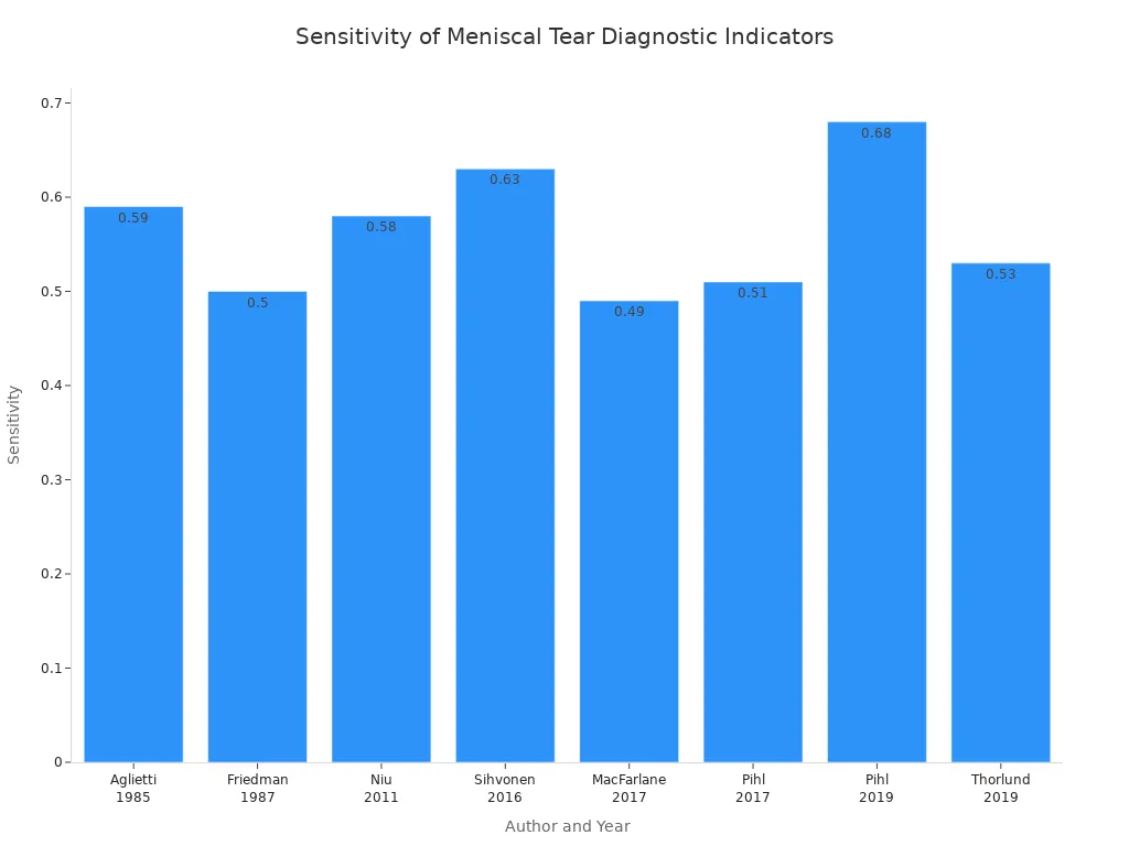
Medial Meniscus Tear: This tear causes occasional “locking” or “catching” of your knee. Your knee might briefly stick or click during movement. Swelling can come and go with certain activities. Twisting or squatting often triggers pain. You feel tenderness along the knee’s joint line. Your knee might unexpectedly ‘give way’. You may also experience stiffness after rest or discomfort during prolonged sitting. MRI is the best imaging test for this. Ultrasound can show your knee in motion. Physical tests like McMurray and Thessaly tests also help.

Pes Anserine Bursitis: This condition causes pain below your knee joint. It often improves with rest. The pain worsens during physical activity or climbing stairs. You feel pain on the inner side of your knee, about 2 to 3 inches below the joint. You might feel discomfort when kneeling, standing from a chair, or walking up or down stairs. Common causes include repetitive knee movements in sports, tight hamstrings, or abnormal knee alignment.
Knee Osteoarthritis: This is a common cause of knee pain. It involves the breakdown of cartilage in your knee. You feel a deep, aching pain on the inner side of your knee. This pain often worsens with activity and improves with rest. Stiffness, especially in the morning, is a common symptom. You might also hear grinding or popping sounds. This type of arthritis can affect one or both knees.
Pain on Outer Knee
Pain on the outer side of your knee often relates to friction or ligament issues.
Iliotibial (IT) Band Syndrome: This causes knee pain on the outside of your knee. The pain worsens with activities that involve repetitive bending and straightening of your knee, like running or cycling. You might feel a clicking, popping, or snapping sensation on the outside of your knee. This happens when the IT band rubs over your thighbone. It leads to inflammation. Risk factors include running on tilted surfaces, sudden increases in training, or worn-out shoes.
Lateral Collateral Ligament (LCL) Injury: You might hear a popping sound at the time of injury. You feel pain or tenderness on the outside of your knee. Swelling, stiffness, and bruising are common. Your knee might feel unstable, like it is locking, buckling, or giving out. A doctor will ask about your injury and symptoms. They will perform a physical exam. Imaging tests like X-rays or MRIs can confirm the diagnosis.
Lateral Meniscus Tear: You feel pain, especially on the outside of your knee. Swelling and stiffness increase after the injury. You might experience a catching sensation or your knee locking. Your knee might feel unstable, like it is “giving away.” You may also find it hard to straighten your knee all the way. Causes include sudden twists or turns when your foot is planted. Older individuals might get tears from simple actions like squatting due to worn cartilage.
Pain Below Kneecap
Pain below your kneecap often involves the patellar tendon or growth plates in adolescents.
Patellar Tendinitis: As mentioned before, this causes acute pain at the bottom edge of your kneecap. You feel pain when you push on the tendon with your knee straight. The pain lessens or disappears when your knee bends to 90 degrees and you push on the tendon.
Osgood-Schlatter Disease: This affects adolescents. It is a repetitive strain injury. You feel knee pain, especially just below your kneecap at the top of your shin. Swelling and tenderness are common. You might have tight quadriceps muscles. A hard, painful bump can form on or just below your kneecap. This pain usually develops gradually. It worsens during and after physical activities like running or jumping.
Pain Behind Knee
Pain behind your knee can indicate issues with fluid buildup, muscles, or the meniscus.
Baker’s Cyst: This is a fluid-filled sac behind your knee. It often results from underlying knee problems like knee arthritis or injuries. You might feel a bump behind your knee. You can experience knee pain, stiffness, or trouble bending your knee. Swelling can extend into your thigh or calf. Many people have no symptoms at all. The swelling might disappear when your knee is bent.
Hamstring Strain: You feel sudden, sharp pain in the back of your thigh, which can extend to behind your knee. This often happens during activities that involve sprinting or sudden stops. You might have bruising and tenderness.
Posterior Meniscus Tear: Similar to other meniscus tears, this can cause pain, swelling, and a catching or locking sensation behind your knee. You might find it hard to fully bend or straighten your knee.
When to Seek Medical Attention for Knee Pain
You need to know when your knee pain requires a doctor’s visit. Some symptoms mean you need immediate care. Other signs suggest you should see a specialist soon. Do not ignore serious knee problems.
Urgent Care Indicators
You should seek immediate medical attention for your knee if you experience certain symptoms. These signs point to a serious injury or condition.
Sudden, Severe Pain: If you have sudden, severe knee pain with no clear cause, get help. This pain might limit your movement. It can also stop you from putting weight on your leg. This could mean a meniscus tear, a torn ligament, a fractured bone, or a dislocated kneecap.
Trauma and Inability to Bear Weight: You need urgent care after an injury like a fall or car accident. This is especially true if you cannot put weight on the affected leg. If you suspect a broken bone, seek help right away. You might also have trouble moving your knee or leg.
Visible Deformity: If your knee looks deformed, you need immediate medical attention. This suggests a serious structural issue.
Signs of Infection: Look for fever, swelling, redness, and warmth around your knee joint. These are signs of a joint infection. You might also have limited range of motion.
Persistent Pain Concerns
Sometimes, knee pain is not an emergency, but it still needs professional evaluation. You should not let knee pain linger.
Pain Lasting More Than a Few Weeks: If your knee pain lasts more than a few weeks, you need a medical evaluation. This is especially important if the pain affects your daily routines or sleep.
No Improvement with Home Remedies: If your pain lasts longer than a week or two without getting better, even with rest, ice, compression, and elevation (RICE), seek help. Your knee needs a doctor’s assessment.
Worsening Symptoms: If your knee pain gets worse over time, you need to see a doctor. Do not wait for it to become unbearable.
Persistent Pain After Surgery: If you have consistent knee pain, swelling, and bruising months after knee surgery, this may indicate a surgical failure. You should consult your surgeon.
Symptoms Requiring Evaluation
Certain symptoms indicate you need a doctor’s evaluation, even if they are not urgent. An orthopedic surgeon can help diagnose these issues.
Swelling and Warmth: Swelling and warmth around your knee joint can mean an inflammatory condition like arthritis. It could also be an infection. You might also have limited range of motion and stiffness.
Instability or “Giving Way”: If your knee feels unstable or buckles, it signals a ligament tear. An ACL tear often causes this sensation of your knee giving way.
Knee Locking or Catching: Your knee might lock, meaning you cannot move it in any direction. Or, movement might be severely limited by pain. This can happen due to ligament tears, dislocation, or fractures. A knee “locked” in flexion, where you cannot extend it, often points to a meniscal tear. You might also experience painful clicking.
Inability to Fully Extend Your Knee: If you cannot fully straighten your knee, you should get it checked. This can be a sign of internal damage.
Recurrent Swelling: If you have swelling that keeps coming back, or persistent pain after trying conservative treatments, an MRI might be necessary.
You should consult an orthopedic surgeon for sudden pain with injury, visible deformity, or inability to bear weight. They can provide an accurate diagnosis and treatment plan for your knee.
Doctor’s Knee Pain Diagnosis

A doctor’s approach to your knee pain diagnosis differs from your self-assessment. They use specific steps to find the exact problem. This helps them create the best treatment plan for your knee.
Clinical Examination
Your doctor starts with a thorough clinical examination. They observe your knee. They look for limping or any bowing or knock-kneed shape. They check for scars, swelling, redness, or muscle loss around your knee. They also feel your knee. They compare your painful knee with your healthy one. They check for warmth, which suggests inflammation. They feel for crepitus, a crackling sound, when your kneecap moves. You will move your knee to show its full range of motion.
Doctors perform specific tests to check your knee’s stability. They test your Anterior Cruciate Ligament (ACL) with the Lachman’s test. This test checks for forward movement of your shin bone. They also test your Posterior Cruciate Ligament (PCL) and collateral ligaments. These tests help them understand your knee’s strength and movement. They also check for meniscus tears using tests like McMurray’s test. This helps pinpoint the source of your knee pain.
Diagnostic Imaging
After the physical exam, your doctor may order imaging tests. These tests help them see inside your knee.
X-rays are often the first step for knee pain. They show signs of fractures, infections, and arthritis. X-rays do not show soft tissues well. For example, they cannot clearly show ligaments or the meniscus. However, X-rays are a good screening test for older patients. They can show if an MRI would be helpful. Dr. Muyibat Adelani says, “X-rays are an appropriate screening test for knee pain in older patients, and often the results of an x-ray can tell whether an MRI would be even helpful.”
An MRI gives a detailed view of your knee’s cartilage, muscles, ligaments, and bones. Doctors use an MRI for acute knee trauma or chronic knee pain when X-rays are not enough. It can find torn ACLs or meniscus tears. An MRI helps when your knee pain lasts longer than three weeks or if you have reduced range of motion.
Treatment Overview
Once your doctor has a clear knee pain diagnosis, they discuss treatment options. Many non-surgical treatments can help your knee. These include physical therapy to strengthen muscles and improve flexibility. Cortisone injections can reduce pain and inflammation. Viscosupplementation injections add a natural gel to your knee. This helps with joint motion. Platelet-rich plasma (PRP) therapy uses your own blood to help tissue repair. Bracing can also provide stability and reduce pain. Lifestyle changes, like weight loss, also help reduce stress on your knee.
This chart helps you understand your knee pain. It gives you initial insights into your discomfort. Remember, this guide is not a definitive diagnosis for your knee. Listen to your body. Prioritize professional medical evaluation for your knee. Doctors provide accurate diagnosis and treatment for your specific knee problem. Effective treatments are available to manage your knee pain and improve your quality of life.