

The large intestine often receives less attention than other digestive organs. However, it performs a crucial task in the digestive system. While the small intestine handles most nutrient absorption, the large intestine has distinct responsibilities. This large intestine absorbs significant amounts of water. For instance, the large intestine reabsorbs almost one liter of fluid daily, preventing its loss. A large intestine diagram helps visualize its structure. Its important role is vital for overall digestive health. This guide explores its anatomy, function, and overall digestive contribution.
Key Takeaways
The large intestine absorbs water and electrolytes. This prevents dehydration and maintains fluid balance.
It forms and stores feces. This prepares waste for elimination from the body.
The large intestine hosts good bacteria. These bacteria help digest food and make important vitamins.
Mucus in the large intestine protects its lining. It also helps move waste smoothly.
A healthy large intestine is important for overall health. It supports the immune system and prevents digestive problems.
Large Intestine Diagram: Anatomy Overview
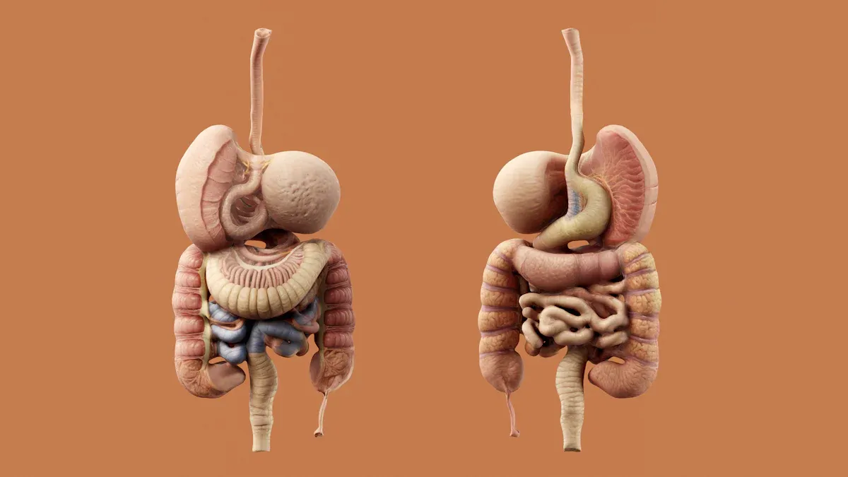
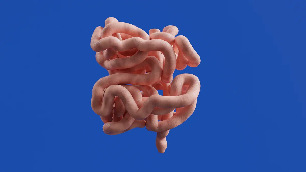
The large intestine, a vital part of the human digestive system, serves as the terminal section of the alimentary canal. It receives digested food from the small intestines. This organ plays a crucial role in the final stages of digestion. The large intestine is approximately 5 feet long. A detailed large intestine diagram helps visualize its complex anatomy and the various parts that contribute to its function. Understanding its anatomy is key to grasping its digestive role.
Main Anatomical Divisions
The large intestine’s anatomy includes several distinct divisions. These parts of the large intestine are defined by specific anatomical landmarks. The cecum, the first part of the large intestine, joins the terminal ileum at the ileocolic junction. The ileocecal valve regulates this junction. The ascending colon extends upwards from the cecum to the right colic (hepatic) flexure. The transverse colon then spans across the abdomen, connecting the right and left colic (splenic) flexures. The descending colon runs downwards from the left colic flexure to the sigmoid colon. The S-shaped sigmoid colon travels from the left iliac fossa to the third sacral vertebra, marking the rectosigmoid junction. The rectum stretches between the rectosigmoid junction and the anal canal. The anal canal forms the terminal part, extending from the anorectal junction to the anus. This entire structure, including the colon and rectum, is a critical component of the gastrointestinal tract.
The Cecum and Appendix
The cecum represents the beginning of the large intestine. It is a pouch-like structure located in the lower right abdomen. The average inner diameter of the cecum in an adult human is 8.7 cm, with a reported range of 8.0 cm to 10.5 cm. The adult cecum measures approximately 7.5 cm in width. Attached to the cecum is the appendix, a small, finger-shaped pouch. The average length of the appendix is 9 centimeters (approximately 3 inches). Its length can vary significantly, from 2 centimeters (about 1 inch) up to 20 centimeters (about 8 inches). The appendix has no known digestive function but contains lymphoid tissue.
Colon Subdivisions
The colon, the longest section of the large intestine, divides into four main parts. Each part has a specific anatomical position and role.
Ascending Colon: This is the first part. It is located on the right side of the abdomen. It extends upwards from the cecum to the right colic flexure. This section measures approximately 5-10 cm long and sits behind the peritoneum.
Transverse Colon: This second segment stretches horizontally across the abdomen from right to left. It is more mobile because the peritoneum surrounds it. The transverse mesocolon supports it.
Descending Colon: This part is found on the left side of the abdomen. It extends downwards from the left colic flexure to the sigmoid colon. Like the ascending colon, it is located behind the peritoneum.
Sigmoid Colon: This is the final S-shaped section. It connects the descending colon to the rectum. The peritoneum surrounds it, allowing for its mobility due to its connection to the pelvic wall via the sigmoid mesocolon.
These subdivisions of the colon are crucial for the movement and processing of waste material. The anatomy of the colon and rectum is complex and vital for digestive health.
The Rectum and Anus
The rectum follows the sigmoid colon. It serves as a temporary storage site for feces before elimination. The rectum is approximately 15 cm in length. It measures approximately 12 to 15 cm in length in the adult. The rectum then transitions into the anal canal. The rectum and anal canal together form the final segment of the large intestine.
The anal canal ends at the anus, the external opening of the gastrointestinal tract. The rectum and anus work together to control defecation. Understanding the function of the rectum and anal canal is important for overall digestive health. The final stages of waste elimination involve the rectum and anal canal.
Internal Structure and Layers
The wall of the large intestine, like other parts of the gastrointestinal tract, consists of four main layers. These layers, from the inner cavity outwards, include the mucosa, submucosa, muscular layer, and serosa/adventitia.
Mucosa: This is the innermost layer. It directly contacts the digested food. It has three sub-layers: the epithelium, lamina propria, and muscularis mucosae. The large intestine’s epithelium lacks villi, unlike the small intestine. Instead, it has a flat surface with thousands of glands. Goblet cells are specialized intestinal epithelial cells within the mucosa. They produce and secrete mucus. This mucus coats the intestinal surface. It helps protect the lining and lubricate the passage of waste.
Submucosa: This layer contains nerves, including the submucous plexus, blood vessels, and elastic fibers with collagen.
Muscular layer: This layer surrounds the submucosa. It comprises two layers of smooth muscle: an inner circular layer and an outer longitudinal layer. In the colon, the outer longitudinal layer thins into three distinct bands called taeniae coli. These bands help form the characteristic pouches (haustra) of the colon.
Serosa/Adventitia: This is the outermost layer. It consists of loose connective tissue. The serosa covers parts of the large intestine within the peritoneum. The adventitia covers retroperitoneal sections.
This detailed anatomy of the large intestine, from its main divisions to its microscopic layers, highlights its complex structure. A large intestine diagram effectively illustrates these intricate components, providing a clear visual understanding of this vital organ. The colon and rectum are essential for the digestive process.
Large Intestine Function: Key Roles
The large intestine performs several vital functions in the body. These functions are crucial for overall digestive health. This section explores the primary roles of the large intestine.
Water and Electrolyte Absorption
The large intestine plays a critical role in absorbing water and electrolytes. It receives chyme, which is mostly liquid, from the small intestine. The large intestine then reclaims much of the remaining water. Water absorption in the large intestine primarily occurs through osmosis. This process operates according to the osmotic gradient. Water also moves passively alongside sodium. The colon largely absorbs this sodium. Aldosterone, a hormone, partly regulates water absorption. It increases sodium absorption in response to volume depletion.
The large intestine primarily absorbs specific electrolytes. These include sodium (Na+) and potassium (K+). Sodium absorption is an active transport process. Mineralocorticoids like aldosterone influence this process. Potassium absorption occurs through a K+-ATPase. This absorption ensures the body maintains proper fluid balance.
Feces Formation and Storage
One major role of the large intestine is to form and store stool. As the large intestine absorbs water and electrolytes, the liquid chyme gradually solidifies. This process creates feces. The large intestine temporarily stores these feces. This storage happens before elimination from the body. Feces are composed of approximately 80% water. The remaining material consists of undigested food, bacteria, and dead cells. The colon’s muscular contractions move this waste material towards the rectum for eventual excretion.
Gut Microbiota and Vitamin Synthesis
The large intestine hosts a vast community of microorganisms. This community is known as the gut microbiota. These bacteria play a significant role in digestion and overall health. The healthy gut microbiota is predominantly constituted by the phyla Firmicutes and Bacteroidetes. Other phyla like Actinobacteria and Verrucomicrobia are also present. The human gut is dominated by two phyla: Bacteroidetes and Firmicutes. Proteobacteria, Fusobacteria, Cyanobacteria, Tenericutes, and Verrucomicrobia are present in minor amounts.
These gut bacteria perform several important functions. They break down complex carbohydrates that human enzymes cannot digest. This process produces short-chain fatty acids. These fatty acids provide energy to colon cells. Gut bacteria also synthesize several essential vitamins. These include various B vitamins:
Biotin
Cobalamin (Vitamin B12)
Folate (Vitamin B9)
Niacin (Vitamin B3)
Pantothenate
Pyridoxine (Vitamin B6)
Riboflavin
Thiamin (Vitamin B1)
For example, bacteria like Bacteroides fragilis and Prevotella copri produce biotin and folate. This vitamin synthesis contributes significantly to the body’s nutrient supply.
Mucus Production
The large intestine produces mucus. This mucus serves several protective functions. It is a complex, dilute, aqueous, and viscoelastic secretion. Its general chemical composition includes a significant amount of water, typically comprising 90–95%. Water acts as a solvent and diffusion medium.
Additionally, mucus contains electrolytes, lipids, and various proteins. The mucus in the large intestine is primarily composed of the gel-forming mucin MUC2. MUC2 forms the structural skeleton of its two layers. These layers include an inner, protective layer that is impermeable to bacteria.
The primary function of mucus is to limit bacterial contact with the epithelium. It also facilitates bacterial transport distally. Intestinal mucus efficiently protects the epithelium. It promotes bacterial clearance and separates bacteria from epithelial cells. This action inhibits inflammation and infection. The inner mucus layer of the colon is particularly important.
It accommodates the large number of gut bacteria. MUC2 mucin is the major component of this mucus system. It forms large net-like polymers that organize the mucus. The stratified mucus layer, along with the glycocalyx of epithelial cells, provides physical protection. It acts as a first line of defense. Mucus forms protective layers in the colon. This prevents direct interaction between bacteria and intestinal tissue. It encapsulates fecal matter and bacteria as they travel through the colon. The gut microbiome promotes protective mucus production.
Digestive Role of the Large Intestine
The large intestine plays a crucial role in the final stages of the digestive system. It processes remaining food material and prepares it for elimination. This organ performs both mechanical and chemical digestion, primarily through bacterial action.
Mechanical Digestion Processes
The large intestine moves its contents using specific muscular contractions. These movements help process the remaining food residue. Haustral contractions are slow, segmented movements. They occur mainly in the transverse and descending colon. A haustrum, when full, contracts and pushes residue into the next haustrum.
These contractions happen about every 30 minutes and last for about one minute. They mix the food residue, which helps with water absorption. Peristalsis in the large intestine is slower than in other parts of the digestive tract. This slow movement allows the colon to absorb most of the water from the feces. Strong waves called mass movements also occur. These waves start in the middle of the transverse colon.
They quickly force contents toward the rectum. Mass movements typically happen three or four times daily, often after eating. The gastrocolic reflex triggers these movements. This reflex responds to stomach distension and digestion byproducts in the small intestine. These movements help move waste out of the colon.
Chemical Digestion by Bacteria
Bacteria in the large intestine perform significant chemical digestion. They break down complex carbohydrates that human enzymes cannot digest. These bacteria ferment various dietary fibers.
Examples include inulin-type fructans, short-chain FOS, oligofructose, and resistant starch. This fermentation produces important end-products. These include acetate, propionate, butyrate, lactate, formate, and succinate. These short-chain fatty acids provide energy to colon cells.
Defecation and Waste Elimination
Defecation is the process of eliminating feces from the body. Neurological reflexes initiate this process. The myenteric defecation reflex increases peristalsis. This moves stool toward the rectum and relaxes the internal anal sphincter.
This reflex is involuntary. The parasympathetic defecation reflex also moves stool. However, individuals can voluntarily control this reflex. The external anal sphincter is a voluntary muscle. People can consciously control it. They can squeeze this muscle to hold a bowel movement. This muscle provides voluntary control for defecation, preventing it until appropriate.
Impact on Overall Digestive Health
A healthy large intestine is vital for overall well-being. The large intestine houses a large population of the body’s lymphocytes. This makes it a major immunological organ.
The gut microbiota helps develop a fully functional immune system. It defends against pathogens by competing for nutrients and secreting antimicrobial peptides. A stable microbiota and mucus layers prevent pathogenic bacterial infections.
Dysbiosis, an imbalance in gut bacteria, negatively affects digestive health. It can cause malnutrition, malabsorption, and food intolerances. Dysbiosis also links to conditions like Irritable Bowel Syndrome (IBS) and inflammatory bowel diseases. It can lead to general digestive difficulties such as diarrhea, constipation, and gas.
The large intestine plays a crucial role beyond just eliminating waste. This vital organ efficiently absorbs water and electrolytes. Its distinct anatomical divisions, clearly shown in a large intestine diagram, facilitate these processes. The large intestine also hosts vital microbial activity, which synthesizes essential vitamins. This organ’s functions are indispensable for overall digestive health. A healthy large intestine is fundamental for well-being and efficient bodily function.
