
The human hand performs countless tasks daily. Its remarkable dexterity allows for intricate movements, from typing to playing musical instruments. The complexity of finger anatomy truly represents a marvel of engineering. This blog post provides a detailed map of the bones, joints, and tendons that make up the finger. Understanding normal hand anatomy reveals how such intricate movements are possible. How does this small part of the hand achieve such incredible precision?
Key Takeaways
- Your fingers have three types of bones: phalanges make up the fingers, metacarpals form the palm, and sesamoid bones act like small pulleys in tendons.
- Finger joints, like your knuckles (MCP joints), allow you to bend, spread, and bring your fingers together for gripping and pinching.
- Tendons help your fingers move; flexor tendons bend them, and extensor tendons straighten them. Ligaments keep your finger joints stable.
- Muscles in your hand (intrinsic) make small, precise movements. Muscles in your forearm (extrinsic) provide strong finger movements.
- Nerves give your fingers feeling and control movement. Blood vessels bring important nourishment to your fingers.
Finger Bones: Skeletal Framework of Finger Anatomy
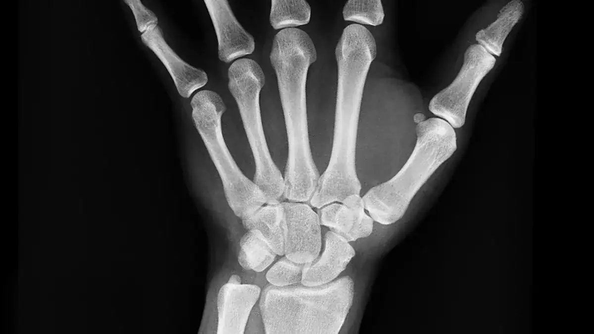
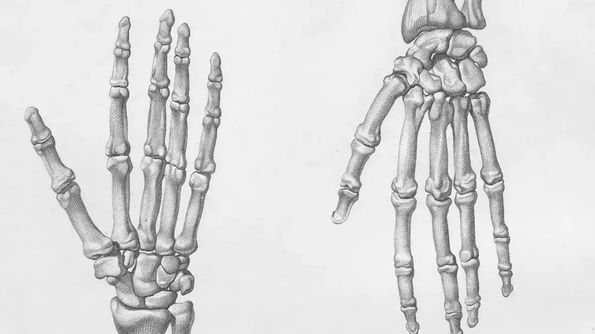
The human hand’s incredible ability to grasp, manipulate, and feel begins with its strong skeletal foundation. The bones of the hand provide the structure for all movements. Understanding the anatomy of the bones of the hand reveals how fingers achieve their remarkable dexterity.
Phalanges: Building Blocks
Phalanges are the individual bones that make up the fingers. They act as the building blocks for each digit. Each finger, except the thumb, contains three phalanx bones. These are the proximal, middle, and distal phalanges. The thumb, however, has only two phalanges: a proximal and a distal phalanx.
Each finger is made up of three long bones called phalanges, except for the thumb, which has only two phalanges.
These bones have specific locations:
- Proximal phalanges: These bones sit closest to the palm of the hand.
- Intermediate (Middle) phalanx: This bone is located between the proximal and distal phalanges. The thumb does not have a middle phalanx.
- Distal phalanges: These bones are found at the very tips of the fingers.
Metacarpals: Hand Connection
Five metacarpal bones form the palm of the hand. These bones connect the phalanges to the wrist bones. Each metacarpal corresponds to a specific finger. For example, the first metacarpal connects to the thumb, and the fifth metacarpal connects to the little finger.
The metacarpal bones create a transverse arch in the hand. This arch provides a stable base for the wrist bones. They also help form the cup shape of the palm. The metacarpals for the thumb and little finger can move more, which helps deepen this cup shape when you bring them together. The index finger’s metacarpal is very stable. The middle metacarpals connect firmly to the wrist. The ring and little finger metacarpals offer more movement, which adds to the hand’s flexibility and grip.
Sesamoid Bones: Small but Significant
Sesamoid bones are small, oval-shaped bones. They resemble sesame seeds. These bones are unique because they are embedded within tendons or muscles, not directly connected to other bones. In the hand, sesamoid bones are often found near joints, especially in the thumb.
Sesamoid bones play an important role. They act like small pulleys. This function provides a smooth surface for tendons to slide over. This action increases the tendon’s efficiency in transmitting muscle forces. They also help protect tendons from too much wear, strain, and injury by redistributing weight and force.
In the thumb, two sesamoid bones are very common at the metacarpophalangeal (MCP) joint. Studies show that almost everyone has these sesamoid bones in their thumb. They are also sometimes present in the MCP joints of the index and little fingers. These small bones are a vital part of the complex finger anatomy.
Finger Joints: Articulations of Finger Anatomy
Joints are crucial parts of the body. They form where two or more bones meet. These connections allow the body to move. The fingers contain several important joints that give them their amazing flexibility. Understanding these joints helps explain the complex movements of the hand.
MCP Joints: The Knuckles
The metacarpophalangeal (MCP) joints are the knuckles. You see them at the base of your fingers. These joints connect the long bones of your hand (metacarpals) to the first bones of your fingers (proximal phalanges). The MCP joint of the thumb is also very important. These joints allow you to bend your fingers, spread them apart, and bring them together. This movement is key for a strong grip and for pinching.
The MCP joints offer a wide range of motion. For the thumb, the average bending (flexion) is about 59.5 degrees, but it can range from 16 to 90 degrees. Straightening (extension) averages 7.9 degrees, with a range from -32 to 58 degrees. Women often show greater flexion and extension in their thumb MCP joints than men. For the other fingers, the MCP joints can extend about 45 degrees. The ability to bend varies, with the little finger often bending more than the index finger.
Several structures keep the MCP joints stable. These structures prevent too much movement and protect the bones.
| Structure | Contribution to Stability The human hand performs countless tasks daily. Its remarkable dexterity allows for intricate movements, from typing to playing musical instruments. The complexity of finger anatomy truly represents a marvel of engineering. This blog post provides a detailed map of the bones, joints, and tendons that make up the finger. Understanding normal hand anatomy reveals how such intricate movements are possible. How does this small part of the hand achieve such incredible precision?
Finger Bones: Skeletal Framework of Finger Anatomy
The human hand’s incredible ability to grasp, manipulate, and feel begins with its strong skeletal foundation. The bones of the hand provide the structure for all movements. Understanding the anatomy of the bones of the hand reveals how fingers achieve their remarkable dexterity.
Phalanges: Building Blocks
Phalanges are the individual bones that make up the fingers. They act as the building blocks for each digit. Each finger, except the thumb, contains three phalanx bones. These are the proximal, middle, and distal phalanges. The thumb, however, has only two phalanges: a proximal and a distal phalanx.
Each finger is made up of three long bones called phalanges, except for the thumb, which has only two phalanges.
These bones have specific locations:
- Proximal phalanges: These bones sit closest to the palm of the hand.
- Intermediate (Middle) phalanx: This bone is located between the proximal and distal phalanges. The thumb does not have a middle phalanx.
- Distal phalanges: These bones are found at the very tips of the fingers.
Metacarpals: Hand Connection
Five metacarpal bones form the palm of the hand. These bones connect the phalanges to the wrist bones. Each metacarpal corresponds to a specific finger. For example, the first metacarpal connects to the thumb, and the fifth metacarpal connects to the little finger.
The metacarpal bones create a transverse arch in the hand. This arch provides a stable base for the wrist bones. They also help form the cup shape of the palm. The metacarpals for the thumb and little finger can move more, which helps deepen this cup shape when you bring them together. The index finger’s metacarpal is very stable. The middle metacarpals connect firmly to the wrist. The ring and little finger metacarpals offer more movement, which adds to the hand’s flexibility and grip.
Sesamoid Bones: Small but Significant
Sesamoid bones are small, oval-shaped bones. They resemble sesame seeds. These bones are unique because they are embedded within tendons or muscles, not directly connected to other bones. In the hand, sesamoid bones are often found near joints, especially in the thumb.
Sesamoid bones play an important role. They act like small pulleys. This function provides a smooth surface for tendons to slide over. This action increases the tendon’s efficiency in transmitting muscle forces. They also help protect tendons from too much wear, strain, and injury by redistributing weight and force.
In the thumb, two sesamoid bones are very common at the metacarpophalangeal (MCP) joint. Studies show that almost everyone has these sesamoid bones in their thumb. They are also sometimes present in the MCP joints of the index and little fingers. These small bones are a vital part of the complex finger anatomy.
Finger Tendons and Ligaments
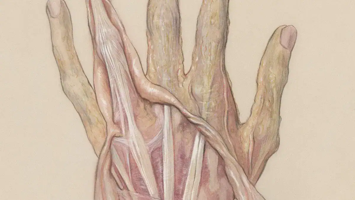
Tendons and ligaments are vital components of the finger’s intricate design. They work together to allow movement and provide stability. Tendons connect muscles to bones. Ligaments connect bones to other bones. This section explores these crucial structures.
Flexor Tendons: Bending
Flexor tendons enable the bending of fingers. When muscles contract, these tendons pull the bones. This action allows for finger movement. The hand contains several important flexor tendons. The Flexor Pollicis Longus tendon specifically controls the thumb’s movement. The thumb’s ability to flex is crucial for many daily activities.
| Tendon Name | Origin | Fingers Flexed |
|---|---|---|
| Flexor digitorum profundus (FDP) | Anterior and medial surfaces of the ulna and the interosseous membrane. | Distal interphalangeal (DIP) joints of the index, middle, ring, and small fingers. |
| Flexor digitorum superficialis (FDS) | Medial epicondyle of the humerus, coronoid process of the ulna, and the radius. | Proximal interphalangeal (PIP) joints of the index, middle, ring, and small fingers. |
| Flexor pollicis longus (FPL) | Anterior surface of the radius and the interosseous membrane. | Interphalangeal (IP) joint of the thumb. |
The Flexor Digitorum Superficialis (FDS) inserts onto the bases of the middle phalanges. It flexes the proximal interphalangeal joints and metacarpophalangeal joints. The FDS also assists with wrist flexion. It can independently flex each finger it connects to. The FDS tendon splits to create an opening. This opening allows the Flexor Digitorum Profundus (FDP) tendon to pass through it. The FDP lies deeper. It flexes the distal interphalangeal joints. These tendons are essential for gripping and fine motor control in the hand. The thumb’s Flexor Pollicis Longus tendon provides strong flexion. This allows the thumb to grasp objects.
Extensor Tendons: Straightening
Extensor tendons work to straighten the fingers. They pull the extensor tendons towards the forearm. This action causes the fingers to extend at the MCP joint. The Extensor Digitorum Communis (EDC) tendons straighten the index, middle, ring, and small fingers. They work with other tendons to extend the fingers at the three finger joints. The Extensor Digiti Minimi (EDM) tendon straightens the small finger. The Extensor Indicis Proprius (EIP) tendon straightens the index finger. These tendons form a complex system for coordinated finger, wrist, and thumb movement. Extrinsic extensor tendons begin in the posterior forearm. They extend into the hand to control wrist and finger movements. The central slip is important for extending the PIP joint. The lateral bands are important for extending the DIP joint. Lumbrical muscles also contribute to smoothing the extension movement. The thumb also has its own extensor tendons, allowing it to extend fully.
Ligaments: Stability and Structure
Ligaments provide crucial stability to the finger joints. They connect bones to other bones. In finger joints, collateral ligaments prevent abnormal sideways bending. The volar plate is another key structure. It forms the floor of the PIP joint. The volar plate prevents hyperextension, which is backward bending, of the PIP joint. Collateral ligaments of the proximal interphalangeal joint (PIPJ) resist ulnar and radial deviation. They have dorsal and volar parts. Dorsal fibers are taut during flexion. Volar fibers are taut during extension. Accessory collateral ligaments help stabilize the volar plate. The volar plate also helps maintain stability of the PIPJ in the anteroposterior plane. It prevents PIPJ hyperextension. This protection is vital for the overall finger anatomy. The MCP joint also has protection against hyperextension by the volar plate. Collateral ligaments protect against radial and ulnar deviation. These structures ensure the hand remains functional and stable. The thumb’s joints also rely on strong ligaments for stability. The thumb’s unique position requires robust ligament support. This support allows the thumb to perform its many tasks.
Finger Muscles, Nerves, Blood Supply
The intricate movements of the fingers rely on a complex network of muscles, nerves, and blood vessels. These systems work together to provide both strength and sensation.
Intrinsic Muscles: Fine Movements
Intrinsic muscles are located entirely within the hand. They allow for fine, precise movements of the fingers. These muscles include the thenar, hypothenar, interossei, and lumbrical muscles. Thenar muscles control the thumb. They sit at the base of the thumb in the palm. Hypothenar muscles line the outer edges of the palm near the pinkie finger. They control that area. Interossei muscles are between the metacarpal bones in the palm. They move fingers side-to-side. Lumbrical muscles are at the base of the four non-thumb fingers. They help bend the fingers. For example, the Opponens Pollicis muscle opposes the thumb. It rotates and flexes the thumb’s metacarpal. Dorsal interossei muscles abduct, or spread, the fingers. Palmar interossei muscles adduct, or bring together, the fingers. Lumbricals flex the metacarpophalangeal joints and extend the interphalangeal joints.
Extrinsic Muscles: Powerful Movers
Extrinsic muscles begin in the forearm. Their tendons extend into the hand. These muscles provide powerful movements for the fingers. Two long flexor muscles are on the underside of the forearm. They control finger bending. The deep flexor connects to the distal phalanx. The superficial flexor connects to the middle phalanx. These are the flexor digitorum profundus and flexor digitorum superficialis. Extensor muscles straighten the fingers. They originate from the humerus and forearm bones. Their bellies are in the forearm. Examples include the extensor digitorum, extensor indicis, and extensor digiti minimi. The extensor pollicis longus and brevis extend the thumb.
Nerves: Sensory and Motor
Nerves carry signals to and from the fingers. They provide both sensation and movement. Three main nerves serve the hand and fingers: the median, ulnar, and radial nerves. The median nerve supplies thenar muscles and the first two lumbricals. It gives sensation to the thumb, index, middle finger, and the radial side of the ring finger. This nerve is key for fine touch and the tripod grip. The ulnar nerve supplies hypothenar muscles, interossei, and the third and fourth lumbricals. It provides sensation to the little finger and the ulnar half of the ring finger. This nerve is important for power grip. The radial nerve is mostly sensory in the hand. It gives protective sensation to the back of the hand and the dorsal side of the thumb, index, middle, and half of the ring finger.
Blood Vessels: Nourishment
Blood vessels form a complex network in the hand. They supply blood to the fingers. The radial and ulnar arteries are the main vessels. They form palmar arches in the hand. The superficial palmar arch and deep palmar arch supply blood. Digital arteries branch from these arches. They carry blood to the digits. Common digital arteries split into proper digital arteries. Each finger has two proper digital arteries. They run along each side. The thumb receives blood from its own digital arteries. This network ensures a constant blood supply to the entire finger anatomy.
The finger’s remarkable function results from an intricate interplay. Bones provide the framework. Joints allow movement. Tendons connect muscles to bones, enabling bending and straightening. Muscles, both intrinsic and extrinsic, power these precise actions. Nerves deliver sensory information and motor commands. Blood vessels ensure constant nourishment. Understanding this detailed anatomy helps people appreciate the hand’s incredible capabilities. It also aids in preventing and rehabilitating injuries.
The human body’s complexity and resilience truly are a marvel of engineering.
FAQ
What are phalanges?
Phalanges are the bones that make up the fingers. Each finger has three phalanges: proximal, middle, and distal. The thumb has two phalanges: proximal and distal. These bones form the finger’s skeletal structure.
What do flexor tendons do?
Flexor tendons allow fingers to bend. Muscles in the forearm connect to these tendons. When these muscles contract, the tendons pull on the finger bones. This action creates the bending motion. They are essential for gripping objects.
What is the purpose of the volar plate?
The volar plate is a strong ligament. It sits on the palm side of finger joints, especially the PIP joint. This structure prevents the finger from bending too far backward. It provides stability and protects the joint from hyperextension.
What is the difference between intrinsic and extrinsic muscles?
Intrinsic muscles are entirely within the hand. They control fine finger movements, like spreading fingers. Extrinsic muscles start in the forearm. Their tendons extend into the hand. They provide powerful movements, such as making a fist.
