
Your heart works tirelessly, a vital pump that sustains your life every moment. It pushes blood throughout your entire body without pause. Did you know a healthy human heart beats approximately 100,000 times daily? This incredible, continuous effort is essential for your overall health.
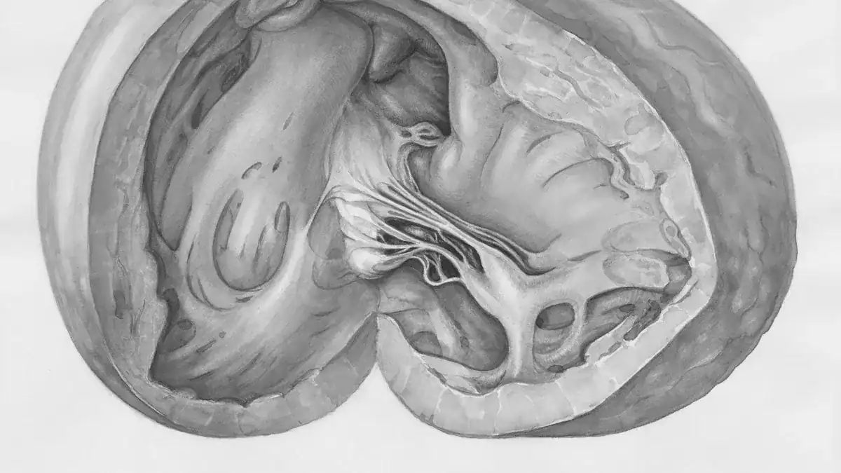
Understanding your heart’s basic anatomy is crucial to appreciating its function. Specifically, its heart chambers and heart valves are key components. These parts ensure proper blood flow. This heart diagram provides a clear map of these essential chambers and valves.
Key Takeaways
Your heart has four chambers. These chambers receive and pump blood.
Your heart has four valves. These valves control the direction of blood flow.
Blood flows through your heart in two main paths. One path goes to your lungs, and the other goes to your body.
The heart wall has three layers. These layers protect the heart and help it pump blood.
Your heart muscle needs its own blood supply. Coronary arteries provide this blood.
Heart’s Location and Overview
Where the Heart Resides
Your heart does not sit directly in the center of your chest. It resides within your thoracic cavity, nestled between your lungs. This specific area is called the mediastinum. Think of the mediastinum as a central compartment. It separates your lungs and contains your heart, major blood vessels, and parts of your esophagus and trachea.
The pericardium, a protective sac, surrounds your heart. This sac separates your heart from other structures within the mediastinum. Your heart’s back surface lies close to your spine. Its front surface sits behind your breastbone and rib cartilage. The top part of your heart, where large blood vessels connect, aligns with your third rib cartilage. The bottom tip, called the apex, points slightly to your left, between your fourth and fifth ribs.
The mediastinum itself has clear boundaries. Your pleurae, which are membranes around your lungs, define its sides. Your chest wall forms its front, and your spine makes up its back. This space extends from your breastbone to your vertebral column. It divides into a superior and an inferior part. The inferior mediastinum, where your heart primarily sits, further subdivides into three regions:
Anterior mediastinum: This area is behind your breastbone and in front of the pericardium.
Middle mediastinum: This central region contains your heart and the beginnings of your great blood vessels.
Posterior mediastinum: This space lies behind the pericardium and in front of your vertebrae.
Basic Heart Structure
Your heart functions as a powerful pump. It continuously moves blood throughout your body. You can think of your heart as a house with strong walls, rooms, and doors. The walls are the muscle tissue. The rooms are the chambers that hold blood. The doors are the valves that control blood flow. Blood vessels act like plumbing pipes, carrying blood away from and back to your heart.
An adult human heart is a surprisingly compact organ. It typically weighs between 250 and 350 grams. This is about the size of your clenched fist. For men, the average weight ranges from 280 to 340 grams. For women, it is usually between 230 and 280 grams. Your heart measures about 5 inches (13 cm) long, 3.5 inches (9 cm) wide, and 2.5 inches (6 cm) thick. This incredible organ’s anatomy allows it to perform its vital work efficiently. This heart diagram will help you visualize these parts.
Heart’s Essential Chambers
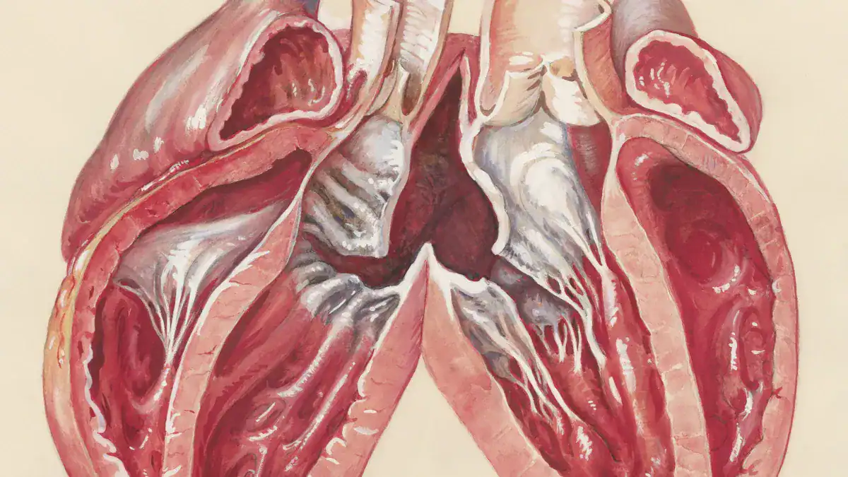
Your heart has four internal spaces, called chambers. These heart chambers work together to move blood. The two upper chambers, the atria, receive incoming blood. The two lower chambers, the ventricles, pump blood out. This heart diagram helps you visualize these crucial parts. Let’s explore each one.
Right Atrium
The right atrium is the first space to receive blood. It collects deoxygenated blood from your body. This blood returns through large veins. The right atrium then sends this blood to the right ventricle. This area has a specific capacity. For women, the calculated right atrial volume is around 20.2 ml/m. For men, it is about 26.1 ml/m. You can also see a wider range of volumes depending on the measurement method:
Measurement Method | Gender | Maximum Right Atrial Volume (mL) | Maximum Right Atrial Volume/BSA (mL/m2) |
|---|---|---|---|
Simpson’s method (RAA included) | Women (adult) | 49-122 | 31-69 |
Simpson’s method (RAA included) | Men (adult) | 59-158 | 32-79 |
Biplane area-length method (RAA excluded) | Women (adult) | 24-81 | 16-54 |
Biplane area-length method (RAA excluded) | Men (adult) | 24-105 | 15-61 |
Inside the right atrium, you find special muscle ridges. These are called pectinate muscles. They are parallel ridges in the atrial walls. In your right atrium, these muscles sit in front of the crista terminalis. The crista terminalis is a strip of muscle. It runs perpendicular to other pectinate muscles. The sinuatrial node, your heart’s natural pacemaker, is near where the crista terminalis meets the superior vena cava opening.
Right Ventricle
The right ventricle is the pumping chamber for deoxygenated blood. It receives blood from the right atrium. Then, it pumps this blood to your lungs. Your lungs add oxygen to the blood. The right ventricle does not need to pump with as much force as the left ventricle. It only sends blood to nearby lungs. The pressure inside your right ventricle shows this. Its systolic pressure typically ranges from 25 to 30 mm Hg. Its end-diastolic pressure is between 5 and 7 mm Hg.
The wall of the right ventricle is also thinner than the left. This reflects its lower pressure workload. You can measure its thickness in different ways:
Measurement Method | Average Thickness (mm) |
|---|---|
RV subcostal (echocardiography) | 6.6 (± 1.6) |
Short-axis method (echocardiography) | 5.5 (± 2.1) |
Autopsy studies show the normal range for both men and women is 0.3–0.5 cm. Echocardiography often shows a range of 0.1–0.5 cm.
Left Atrium
The left atrium receives oxygenated blood. This blood comes directly from your lungs. It enters the left atrium through specific vessels. Four pulmonary veins connect to the left atrium. Their openings are typically in the upper back and side of the atrial wall. Sometimes, the left set of pulmonary veins might join into one common tube.
The left atrium also has pectinate muscles. You find these within the left atrial auricle. This auricle is a highly textured part of the left atrium. After receiving this fresh, oxygen-rich blood, the left atrium sends it to the left ventricle.
Left Ventricle
The left ventricle is the strongest pumping chamber of your heart. It receives oxygenated blood from the left atrium. Then, it pumps this blood out to your entire body. This includes your brain, muscles, and all other organs. The left ventricle must generate high pressure. It pushes blood through a vast network of arteries. Its muscular wall is the thickest of all the heart’s chambers. This powerful pump ensures every part of your body gets the oxygen and nutrients it needs.
Crucial Heart Valves
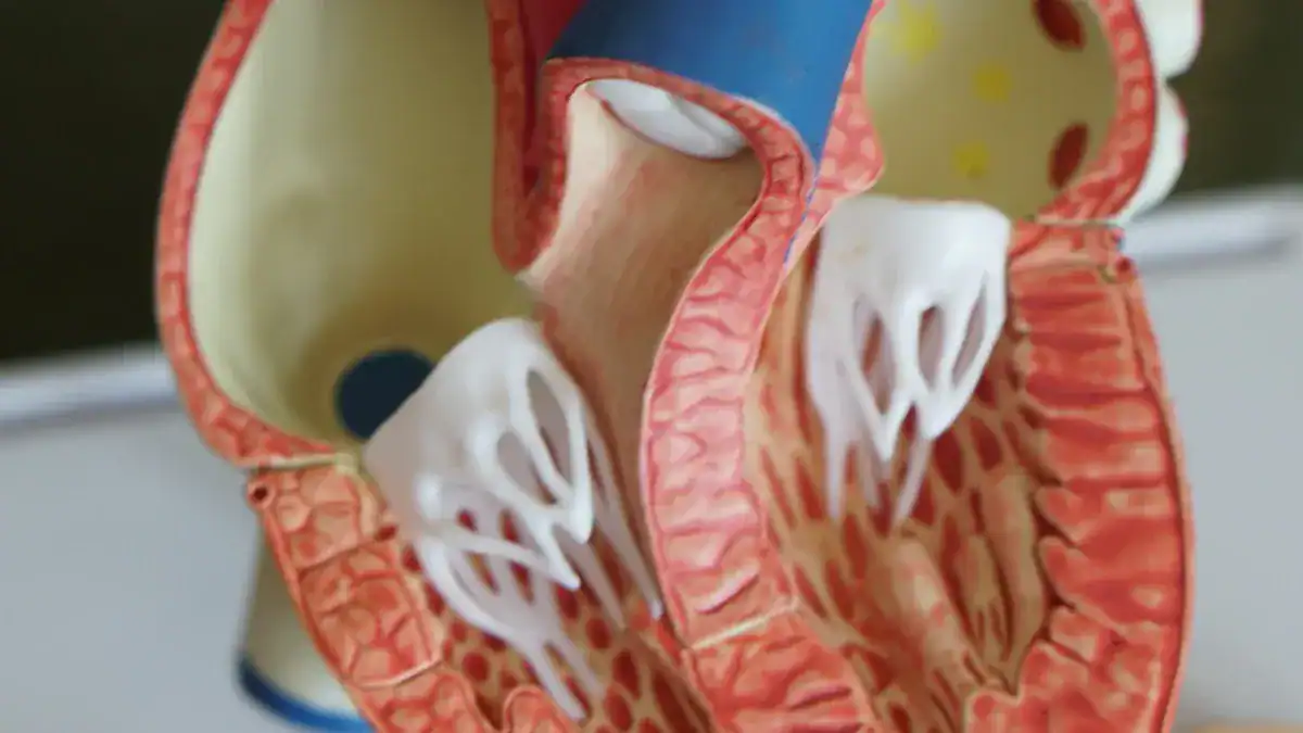
Your heart has four special gates. These are your heart valves. They open to let blood flow through. Then, they close tightly to stop blood from going backward. Think of them as one-way doors. They make sure your blood always moves in the right direction. This is crucial for your heart to pump efficiently.
Atrioventricular Valves
You have two main types of heart valves. The first type is the atrioventricular (AV) valves. These valves sit between your atria and ventricles. They control blood flow from the upper chambers to the lower chambers.
One of these is the tricuspid valve. It separates your right atrium from your right ventricle. The tricuspid valve has three leaflets. These are like small flaps. The base of each leaflet connects to a strong, fibrous ring. Thin cords, called chordae tendineae, attach to the edges of these leaflets. These cords connect to papillary muscles inside your right ventricle. You have three papillary muscles in your right ventricle that support the tricuspid valve.
The other AV valve is the mitral valve. It sits between your left atrium and left ventricle. The mitral valve has two leaflets. You might also hear it called the bicuspid valve. Its leaflets also connect to a fibrous ring at their base. Chordae tendineae attach to the free edges of these leaflets. These cords connect to two papillary muscles in your left ventricle.
Semilunar Valves
The second type of valves is the semilunar valves. These valves control blood flow out of your ventricles. They let blood go into the large arteries that leave your heart.
One semilunar valve is the pulmonary valve. It sits between your right ventricle and the pulmonary artery. This artery carries blood to your lungs. The pulmonary valve has three leaflets. These are also called cusps or flaps. They include an anterior cusp, a left cusp, and a right cusp. These cusps attach to a tough, fibrous ring called the annulus.
The other semilunar valve is the aortic valve. It sits between your left ventricle and the aorta. The aorta is the main artery. It carries oxygen-rich blood to your body. The aortic valve is similar to the pulmonary valve. It also has leaflets.
Unidirectional Flow
Your heart valves work together to ensure blood flows in only one direction. This ensures unidirectional flow.
During systole, your ventricles contract. Your AV valves close. This stops blood from flowing back into your atria. At the same time, your semilunar valves open. This lets blood leave your ventricles. It goes into the pulmonary artery and aorta.
During diastole, your ventricles relax. Your semilunar valves close. This prevents blood from returning to your heart from the arteries. Then, your AV valves open. This allows blood to fill your ventricles from your atria.
The chordae tendineae and papillary muscles help your AV valves work correctly. They prevent the valve leaflets from turning inside out when your ventricles contract.
Your valve leaflets have a special structure. They have three layers of tissue. One layer is rich in elastin. Another layer has many proteoglycans. The third layer is rich in collagen. These layers, along with special cells, make sure your valves are strong and flexible. This layered design is key for keeping blood flowing in one direction.
Sometimes, problems can affect your heart valves. A leaky valve, also called regurgitation, happens when a valve does not close completely. This causes blood to flow backward. For example, in mitral valve regurgitation, blood flows back into the left atrium. This forces your heart to work harder.
Another problem is stenosis. This happens when a valve opening becomes too narrow. The leaflets might thicken or stiffen. This restricts blood flow. Your heart then has to pump much harder to push blood through the narrow opening. Both regurgitation and stenosis can make your heart less efficient.
Pathway of Blood Flow
Your heart works like a powerful, two-sided pump. It sends blood on two main journeys. These journeys are called circulation. One journey goes to your lungs. The other journey goes to the rest of your body. Understanding these paths helps you see how your heart keeps you alive. This heart diagram shows you the route.
Pulmonary Circulation
Pulmonary circulation is the first journey for your blood. It starts when your right ventricle pumps deoxygenated blood. This blood has little oxygen. It travels through the pulmonary artery to your lungs. In your lungs, something amazing happens. Your blood picks up fresh oxygen and releases carbon dioxide.
In your lungs, tiny air sacs called alveoli meet very small blood vessels. These are pulmonary capillaries. Here, gas exchange occurs. This process is vital for your body:
Oxygen moves from the alveoli into your blood. It mostly attaches to hemoglobin in red blood cells. A small amount dissolves in plasma.
Carbon dioxide leaves your blood and goes into the alveoli. You then breathe it out. Some carbon dioxide travels on hemoglobin. Some dissolves in plasma. Some changes form for transport.
This exchange happens through simple diffusion. Gases move from an area of high pressure to an area of low pressure.
After this exchange, your blood is rich with oxygen. It then flows from your lungs back to your heart. It travels through the pulmonary veins. These veins carry the oxygenated blood to your left atrium.
Blood moves quickly through your pulmonary circulation. Scientists measure this transit time. Here is how fast blood typically moves:
Measurement Type | Average Time (seconds) |
|---|---|
Mean Transit Time | 4.67 ± 1.7 |
Median Time | 4.47 ± 1.74 |
Peak Time | 3.98 ± 1.7 |
Onset Time | 2.27 ± 1.43 |
This chart shows the average time blood spends in your pulmonary circulation:
Systemic Circulation
Systemic circulation is the second, larger journey for your blood. It begins when your left ventricle pumps oxygenated blood. This blood is full of oxygen. It leaves your heart through the aorta. The aorta is your body’s largest artery. It branches into many smaller blood vessels. These vessels carry blood to every part of your body. This includes your brain, muscles, and organs.
When blood reaches your body’s tissues, it enters tiny capillaries. Here, your blood delivers oxygen and nutrients to your cells. It also picks up waste products like carbon dioxide. This exchange is complex and happens in several ways:
Diffusion: Small molecules like gases, fats, and fat-soluble substances pass directly through cell membranes.
Facilitated Diffusion: Glucose, amino acids, and ions (like sodium, potassium, calcium, and chloride) use special transporters and channels to cross membranes.
Intercellular Clefts: Glucose, ions, and larger molecules can leave your blood through small gaps between cells.
Fenestrated Capillaries: These capillaries have pores. Larger molecules can pass through them.
Sinusoids: These capillaries have very large gaps. Even big plasma proteins can move through them.
Endocytosis and Exocytosis: Some large plasma proteins move into and out of cells inside small sacs called vesicles.
Osmosis: Water moves across capillary walls.
Bulk Flow (Filtration and Reabsorption): Fluids move in large amounts. Filtration pushes fluid out of capillaries. Reabsorption pulls fluid back into capillaries.
After delivering oxygen and nutrients, your blood becomes deoxygenated. It now carries waste products. This deoxygenated blood returns to your heart. It travels through veins. Smaller veins join to form larger veins. Eventually, two large veins, the superior and inferior vena cava, carry this blood back to your right atrium.
Chambers and Valves in Flow
Let’s trace the complete path of blood through your heart and body. This shows how your heart chambers and heart valves work together.
Deoxygenated blood from your body enters your right atrium.
It passes through the tricuspid valve into your right ventricle.
Your right ventricle pumps this blood through the pulmonary valve into the pulmonary artery.
The pulmonary artery carries blood to your lungs for oxygen.
Oxygenated blood returns from your lungs through pulmonary veins. It enters your left atrium.
It passes through the mitral valve into your left ventricle.
Your left ventricle pumps this oxygenated blood through the aortic valve into the aorta.
The aorta distributes blood to your entire body.
After delivering oxygen, deoxygenated blood returns to your right atrium. The cycle then begins again.
Each valve opens and closes at just the right time. This ensures blood flows in one direction. It prevents any backflow. This precise coordination of chambers and valves makes your heart an incredibly efficient pump.
Supporting Structures
Your heart has important supporting structures. These parts help it work correctly. They protect your heart and keep it healthy.
Heart Wall Layers
Your heart wall has three distinct layers. Each layer plays a vital role in your heart’s function.
Epicardium: This is the outermost layer. It is a thin layer of connective tissue and fat. It protects your heart from trauma or friction. This layer also contains the coronary blood vessels. These blood vessels supply oxygen to your heart tissues. It also secretes fluid to reduce friction during heart contractions.
Myocardium: This is the middle layer. It is the muscle tissue of your heart. Cardiac muscle cells make up this layer. It is the thickest layer. This layer is responsible for your heart’s powerful contractions. It pumps blood throughout your body.
Endocardium: This is the innermost layer. It has endothelial cells. It provides a smooth surface inside your heart. This smooth surface helps blood flow easily. It prevents blood from sticking. This layer also helps regulate how your heart muscle contracts.
Cardiac Septa
Your heart has internal walls called septa. These walls divide your heart into different sections. They keep oxygen-rich blood separate from oxygen-poor blood.
The interatrial septum divides your two upper chambers, the atria. It has an oval-shaped depression called the oval fossa. A raised muscular rim surrounds this fossa. This septum helps keep blood from mixing between your atria.
The interventricular septum divides your two lower chambers, the ventricles. It has two main parts. The lower part is thick and muscular. The upper part is thin and smooth. This septum is crucial. It keeps oxygenated blood in your left ventricle separate from deoxygenated blood in your right ventricle. Sometimes, babies are born with holes in these septa. These are called septal defects. An atrial septal defect is a hole in the wall between the upper chambers. A ventricular septal defect is a hole in the wall between the lower chambers. These holes can cause blood to flow where it should not. This makes your heart and lungs work harder.
Coronary Circulation
Your heart muscle needs its own supply of oxygen-rich blood. The coronary circulation provides this. It is a network of specialized blood vessels.
Coronary arteries deliver oxygenated blood to your heart muscle. The Right Coronary Artery (RCA) supplies the right side of your heart. It also supplies parts of both ventricles. The Left Coronary Artery (LCA) is also called the Left Main Coronary Artery. It branches into two main arteries. These are the Left Circumflex Artery (LCX) and the Left Anterior Descending Artery (LAD). These arteries supply the left side of your heart.
Cardiac veins collect deoxygenated blood from your heart muscle. They return this blood to your right atrium. The Great Cardiac Vein, Middle Cardiac Vein, and Small Cardiac Vein are important examples. Most of these veins drain into a large vessel called the coronary sinus. The coronary sinus then empties into your right atrium. Some smaller veins drain directly into your right atrium. This system ensures your heart muscle always receives the oxygen it needs to pump blood effectively.
Your human heart is a truly sophisticated pump. Its four chambers and precise valves work in perfect coordination. This ensures efficient blood circulation throughout your entire cardiovascular system. Remember, the strong septa and the thick left ventricular myocardium are crucial for this power. Coronary arteries also keep your heart muscle healthy. Understanding this basic anatomy is fundamental. It helps you appreciate your heart health and the intricate design of your body. ❤️
Chambers: Receive and pump blood.
Valves: Control blood flow direction.
Blood Flow: Delivers oxygen and nutrients.
FAQ
What do your heart valves do?
Your heart has four valves. These valves act like one-way doors. They open to let blood flow forward. Then, they close tightly to stop blood from flowing backward. This ensures your blood moves in the correct direction through your heart. Proper valve function is key for good heart health.
What is high blood pressure?
High blood pressure means your blood pushes too hard against your blood vessels. This makes your heart work harder. Over time, high blood pressure can damage your heart and blood vessels. It increases your risk for heart disease and heart attack. You should monitor your blood pressure regularly for good health.
What is a normal heart rate?
Your heart rate is how many times your heart beats per minute. For most adults, a normal resting heart rate is between 60 and 100 beats per minute. Many factors affect your heart rate, like activity and stress. A healthy heart rate shows your heart is working well.
What is a heart attack?
A heart attack happens when blood flow to a part of your heart muscle stops. This usually occurs because a blood clot blocks a coronary artery. Without blood, that part of your heart muscle starts to die. Recognizing symptoms and getting quick medical help is vital for your health.
What is the difference between arteries and veins?
Arteries carry oxygen-rich blood away from your heart to your body. Veins carry oxygen-poor blood back to your heart. Both are types of blood vessels. Your heart pumps blood through these vessels to deliver oxygen and nutrients everywhere.
